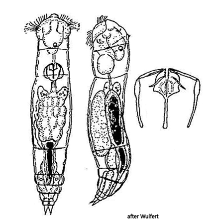
cerebral ganglion rectangular with hemispherical, granular sac
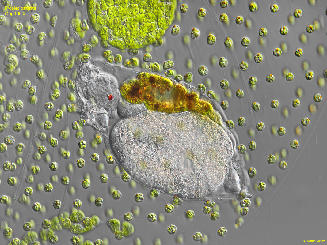
stomach filled with greenish, yellowish and reddish masses
parasitic lifestyle in Volvox, Uroglena and Ophrydium
Proales parasita
I could find Proales parasita only once in June 2017. The identification is not difficult due to the parasitic lifestyle in colonies of Volvox, Uroglena or Ophrydium. The two other parasitic rotifers that live in Volvox or Uroglena are Ascomorphella volvocicola and Cephalodella edax. The first species has no toes and Cephalodella edax has the eyespot on top of the forehead. These features mean that these two species can be excluded.
The specimens in my population were about 10 % larger than the maximum stated length (160 µm). However, there are few independent descriptions of Proales parasita, so little is known about the variability in size. As in many Proales species, the singular eyespot is asymmetrically arranged in the body and shifted to the right side of the body (s. fig. 2). Due to the rich food supply from the cells of their host, the specimens can be opaque and deformed.
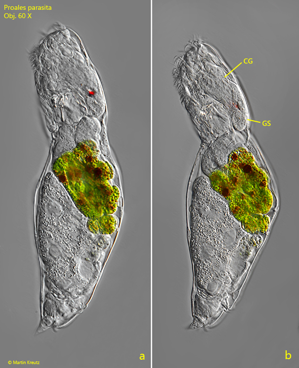
Fig. 1 a-b:Proales parasita. L = 190 µm. Two focal planes of a freely swimming specimen from left. Note the cerebral ganglion with the adjacent granular sac (GS). Obj. 60 X.
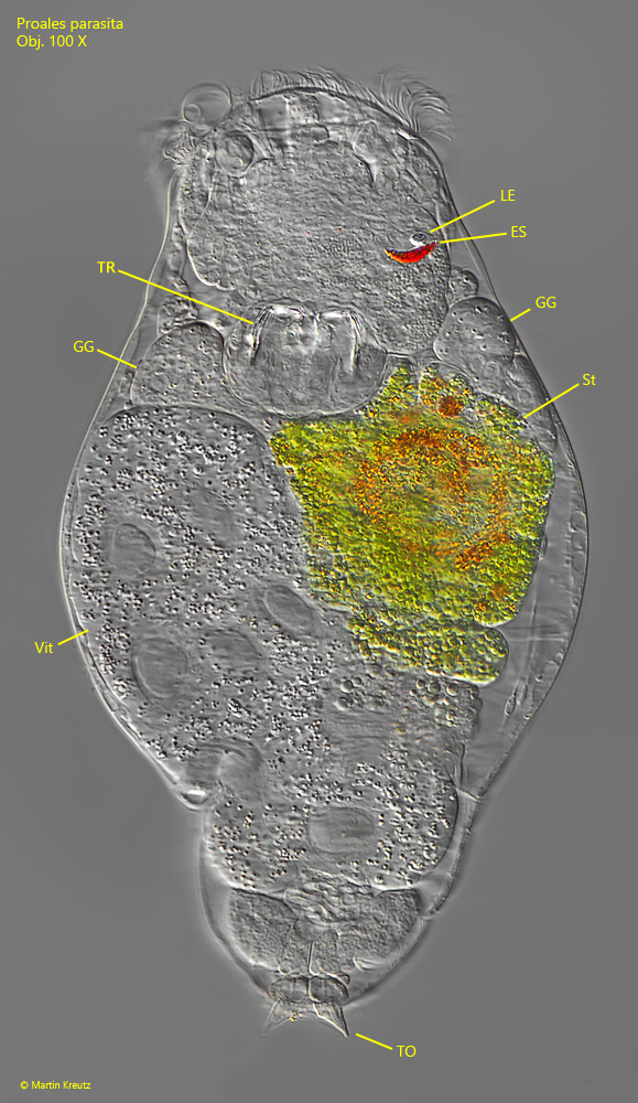
Fig. 2:Proales parasita. A squashed specimen from dorsal. Note the eyespot (ES) with a lens (LE) shifted to the right side. GG = gastric glands, ST = stomach, TO = toes, TR = trophi, Vit = vitellarium. Obj. 100 X.
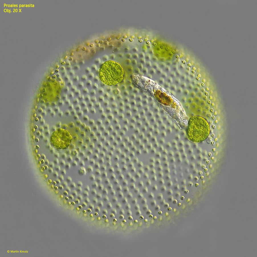
Fig. 3:Proales parasita. L = 180 µm. A specimen parasitizes in a colony of Volvox aureus and feeds on the host’s cells. Obj. 100 X.
Fig. 4:Proales parasita. The specimen as shown in fig. 3 in detail. Obj. 100 X.
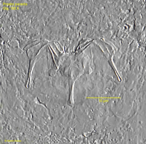
Fig. 5:Proales parasita. The trophi in a strongly squashed specimen. Obj. 100 X.