body elongated spindle-shaped, head clearly set off
dorsal side curved, ventral flat
length 125–268 µm
corona reduced, ventrally shifted
cuticle clear, transparent, covered with mucilaginous sheath
mucilaginous sheath often interspersed with bacteria
stomach and intestine clearly separated
lateral antennae with long setae
dorsal antenna and caudal antenna on a distinct papilla
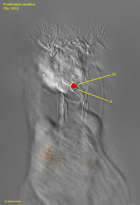
one eyespot with lens
toes 16–22 µm long, narrow and pointed
two distinct pedal glands
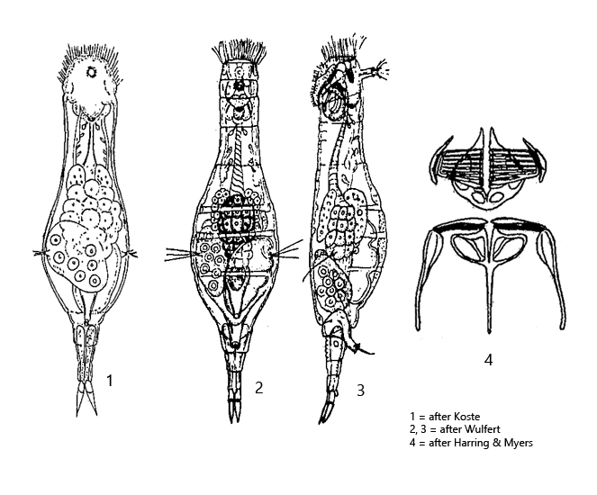
Proalinopsis caudatus
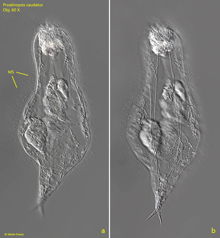
I find Proalinopsis caudata regularly in the Simmelried, especially in old samples with decaying plants. This rotifer has a mucilaginous sheath (s. figs. 3 b, 4 a and 5), which can be seen well especially in low magnifications. The head is clearly separated from the body (s. fig. 1 a-b), which can be seen especially well in lateral view. The locomotion is a slow gliding.
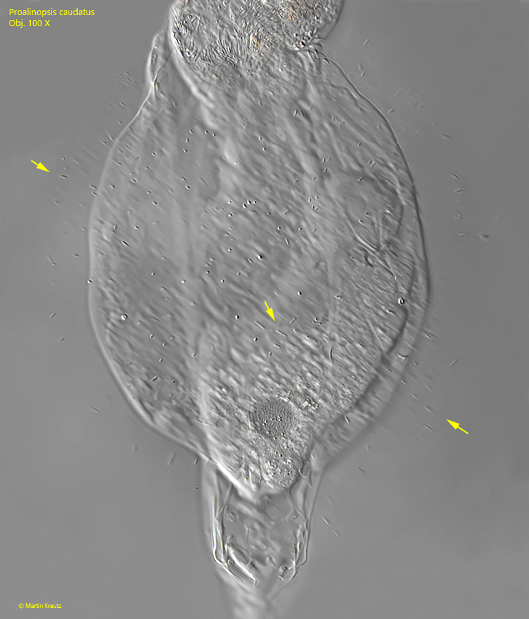
The mucilaginous sheath is interspersed with bacteria. However, these are not interspersed randomly, but are radially oriented and are mostly located in the outer layer of the mucilaginous sheath. In my population, the bacteria in the sheath were 2–4 µm long rods (s. fig. 5). Whether the bacteria use the excreted mucilaginous sheath as a food source, or whether they also provide a benefit to Proalinopsis caudatus, cannot be readily determined. However, it is striking that they are obviously bacteria of the same species and are aligned along the growth structure of the mucilaginous sheath.
The large rotifer Notommata copeus has also got a mucilaginous sheath that is interspersed with bacteria in the same manner. This rotifer also moves only by slowly gliding and is often found between floating plant masses. Obviously the presence of a mucilaginous sheath offers an advantage for this lifestyle, which remains unresolved for the time being.
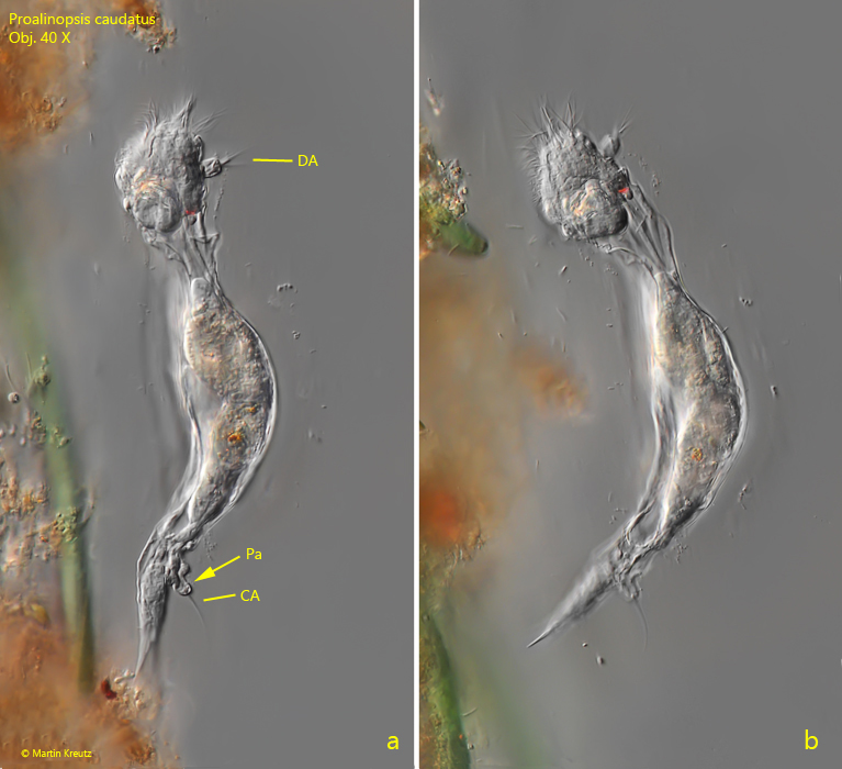
Fig. 1 a-b:Proalinopsis caudatus. L = 180 µm. A specimen gliding along a detritus flake from left lateral view. Note the dorsal antenna (DA) and the caudal antenna (CA) arising from short papillae. The head is clearly set off from the body. Obj. 40 X.
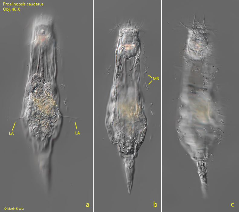
Fig. 2 a-c:Proalinopsis caudatus. L = 217 µm. A freely gliding specimen from dorsal. Note the lateral antennae with the long setae. The body is covered with a mucilaginous sheath (MS), visible by interspersed bacteria. Obj. 40 X.
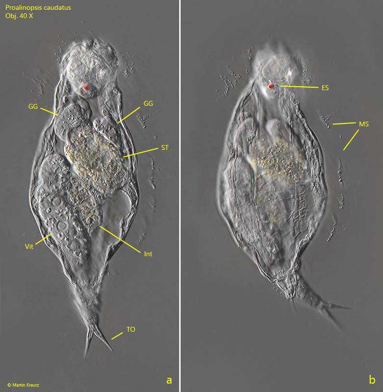
Fig. 3 a-b:Proalinopsis caudatus. L = 217 µm. The slightly squashed specimen shown in fig. 2 a-c. ES = eyespot, GG = gastric glands, Int = intestine, MS = mucilaginous sheath, ST = stomach, TO = toes, Vit = vitellarium. Obj. 40 X.
Fig. 4 a-b:Proalinopsis caudatus. L = 216 µm. A transparent, slightly squashed specimen from ventral. Note the mucilaginous sheat (MS) with the interspersed bacteria. Obj. 60 X.
Fig. 5:Proalinopsis caudatus. L = 216 µm. Focal plane on the surface of the mucilaginous sheath with the interspersed bacteria (arrows). The bacteria are 2–4 µm long rods and arranged radially. Obj. 100 X.
Fig. 6 a-b:Proalinopsis caudatus. L = 140 µm. A freely gliding specimen from ventral. Co = corona, PG = pedal glands. Obj. 60 X.
Fig. 7:Proalinopsis caudatus. A slightly squashed specimen with focal plane on the eyespot (ES) with an attached lens (LE). Obj. 100 X.
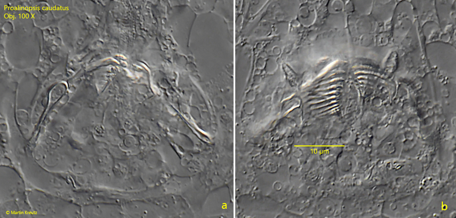
Fig. 8 a-b:Proalinopsis caudatus. Two focal planes of the trophi in a strongly squashed specimen. Obj. 100 X.