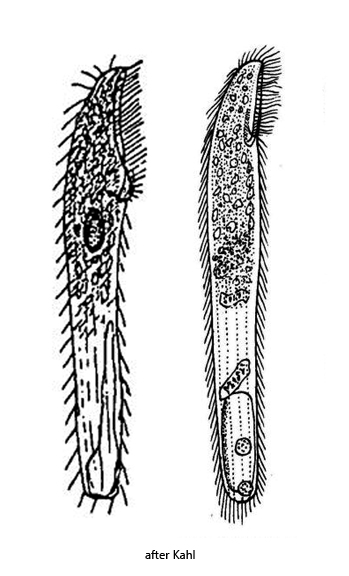
body elongated, parallel-sided, posterior end bluntly rounded
length 100–200 µm, width 10–25 µm
cytoplasm appears pink by hundrets of symbiotic purple sulfur bacteria
some symbiotic algae are scattered between the purple sulfur bacteria
adoral zone reaching to anterior sixth or fourth before it bent to the right
to right side of adoral zone a undulating membrane of short cilia
5–6 ciliary rows per side
between the rows of cilia stripes of refractive granules
macronucleus ellipsoid, near mid-body
several micronuclei adjacent to the macronucleus
contractile vacuole large, terminal, without canal
Pseudoblepharisma tenue
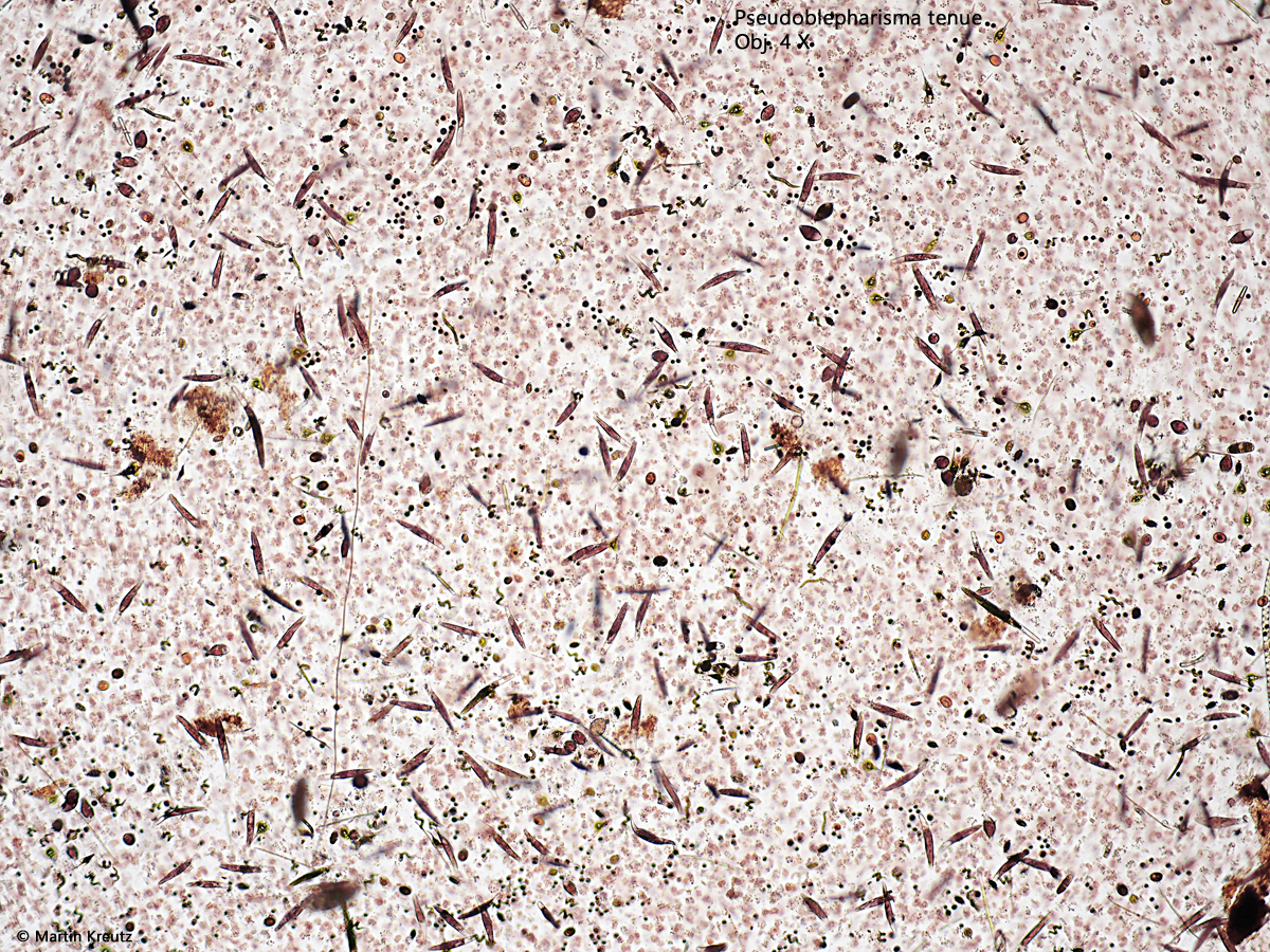
I find Pseudoblepharisma tenue exclusively in the Simmelried. There it is one of the most common ciliates and sometimes emerges in masses (s. fig. 1). This is remarkable because Kahl and later authors have found Pseudoblepharisma tenue only rarely and sporadically. Pseudoblepharisma tenue is particularly common in layers with a high concentration of rhodobacteria. This is the case in areas where dead plant masses decompose anaerobically.
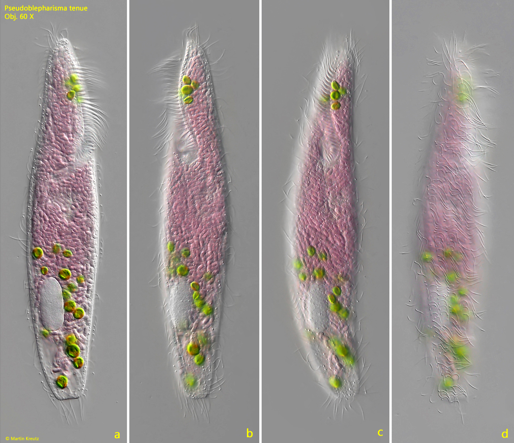
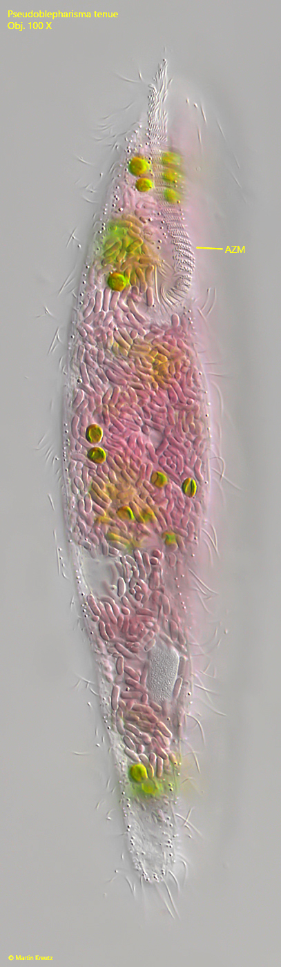
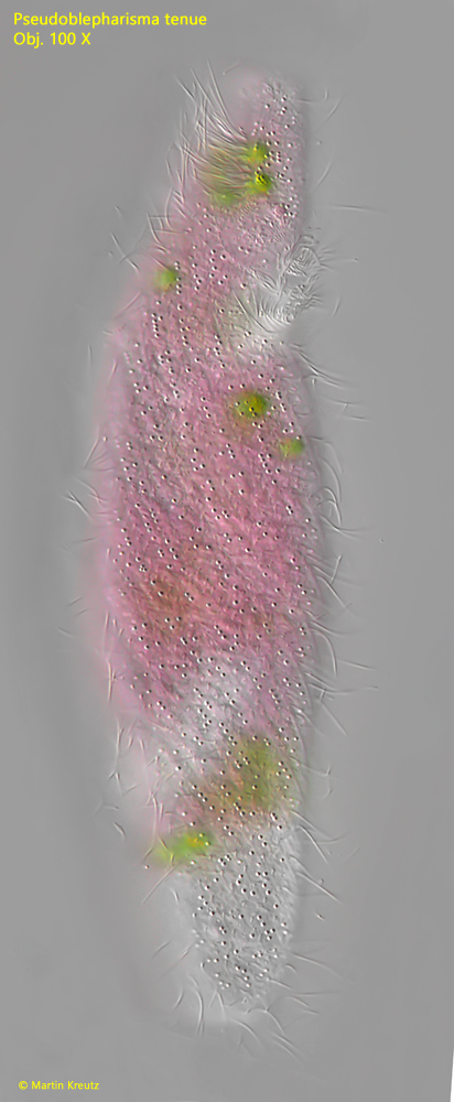
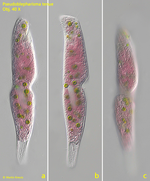
Pseudoblepharisma tenue resembles Spirostomum in body shape. The shape is slender with an adoral zone, which reaches to the first quarter (s. fig. 3). There are 5–6 rows of cilia on each side of the body, as described by Kahl (s. fig. 2 d). Between the rows of cilia are stripes of granules inserted (s. fig. 4). The macronucleus is ellipsoid with several adjacent micronuclei, which are lenticular (s. fig. 5). The most striking feature, however, is the intense purple color, which is caused by hundreds of symbiotic purple sulfur bacteria in the cytoplasm. According to Kahl, the purple sulfur bacteria should be concentrated in the anterior half of the cell. In my population, however, the cells were always completely filled with purple sulfur bacteria, except for the terminal area were the contractile vacuole is located (s. fig. 2 a-d). In addition to the purple sulfur bacteria, there are always about 20 symbiotic algae in the cytoplasm, which are interspersed between the bacteria. I have never found a specimen without these symbiotic algae.
Only very few ciliates are known to have purple sulfur bacteria as symbionts. However, a combination with algae as symbionts is unique. Both symbionts actually have different habitat requirements. Purple sulfur bacteria prefer an anaerobic environment and algae an aerobic one. How Pseudoblepharisma tenue regulates the coexistence of these contrasting symbionts and what benefits the ciliate derives from this was investigated in detail by Muñoz-Gómez et al. (2021).
As a result of the study, it was shown that the purple sulfur bacteria belong to the genus Thiodictyon, but have a genome that has been reduced by half. As a result, for example, the ability to use reduced sulphur compounds as electron donors has been lost. The symbiotic algae belong to the genus Chlorella and have mitochondria for aerobic respiration. The photosystems of the purple sulfur bacteria absorb between 700–900 nm and those of the algae between 400–700 µm. The metabolic products of the two symbionts and the host are used by each other, like a kind of ring exchange. Due to the different abilities of its symbionts, Pseudoblepharisma tenue is able to colonize both light aerobic and dark anaerobic habitats. However, the high population of purple sulfur bacteria in the plasma indicates that Pseudoblepharisma tenue prefers anaerobic habitats. The two symbionts therefore give the ciliate greater flexibility and adaptability.
Fig. 1:Pseudoblepharisma tenue. A mass development in a sample with a high concentration of colonies of purple bacteria. Obj. 4 X.
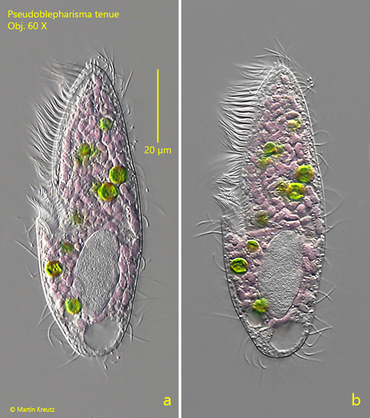
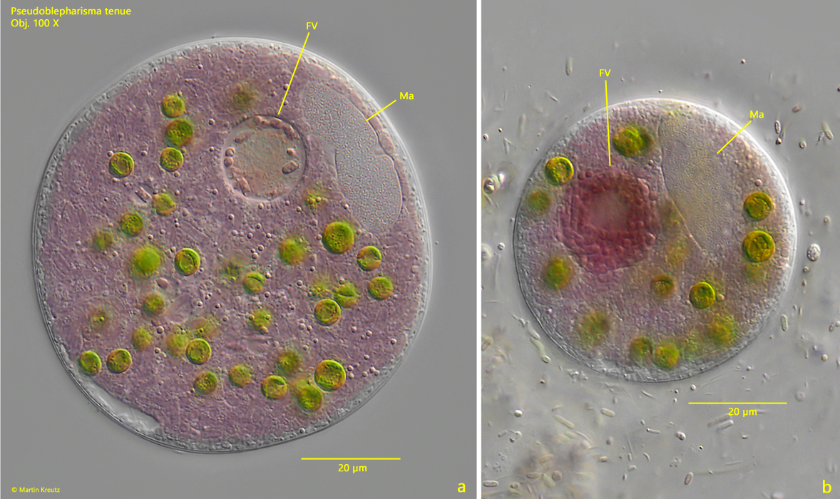
Fig. 2 a-d:Pseudoblepharisma tenue. L = 175 µm. Different focal planes of a freely swimming specimen. Note the intense purple color caused by the symbiotic purple sulfur bacteria in the cytoplasm. Obj. 60 X.
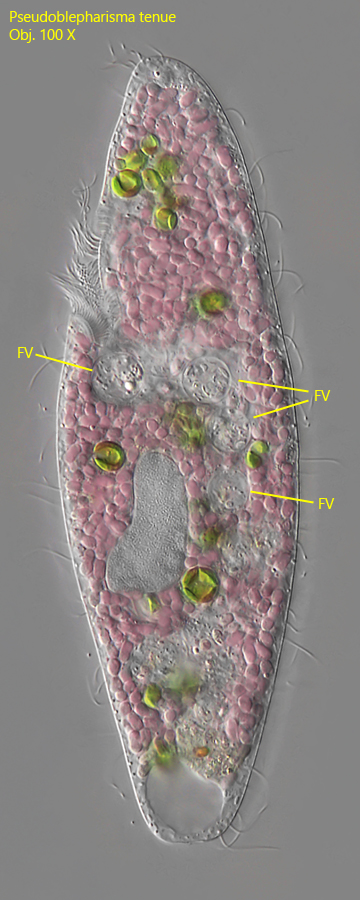
Fig. 3:Pseudoblepharisma tenue. L = 182 µm. Ventral view of a slightly squashed specimen. Note the adoral zone of membranelles (AZM) bent to the right into the mouth opening. Obj. 100 X.
Fig. 4:Pseudoblepharisma tenue. Focal plane on the rows of refractive granules arranged between the ciliary rows. The granules are colorless and have a diameter of 0.6 µm. Obj. 100 X.
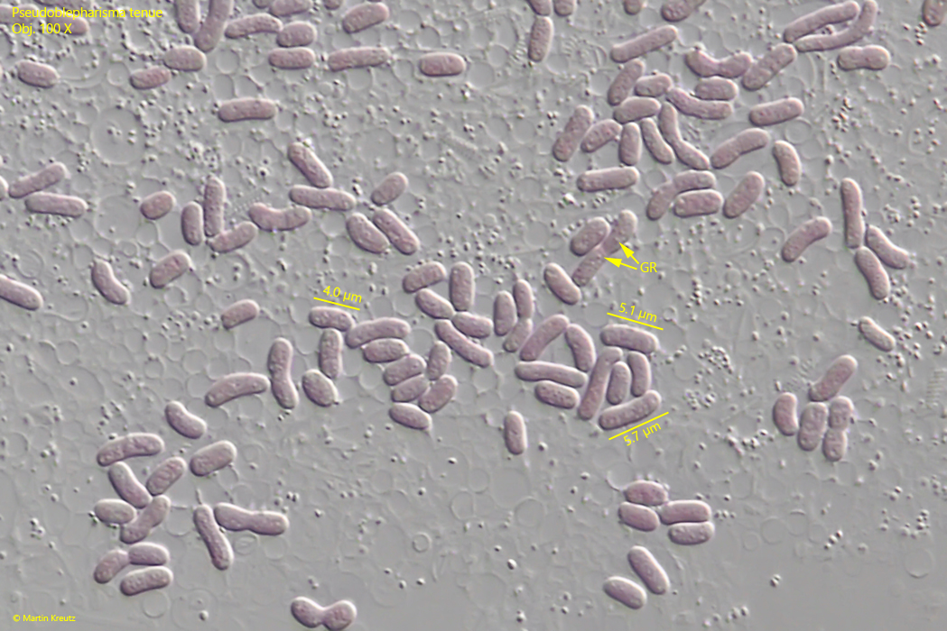
Fig. 5:Pseudoblepharisma tenue. The symbiotic purple sulfur bacteria in a strongly squashed specimen. Non dividing bacteria are about 4–6 µm long. Note the fine granules (GR) in the cells. Obj. 100 X.
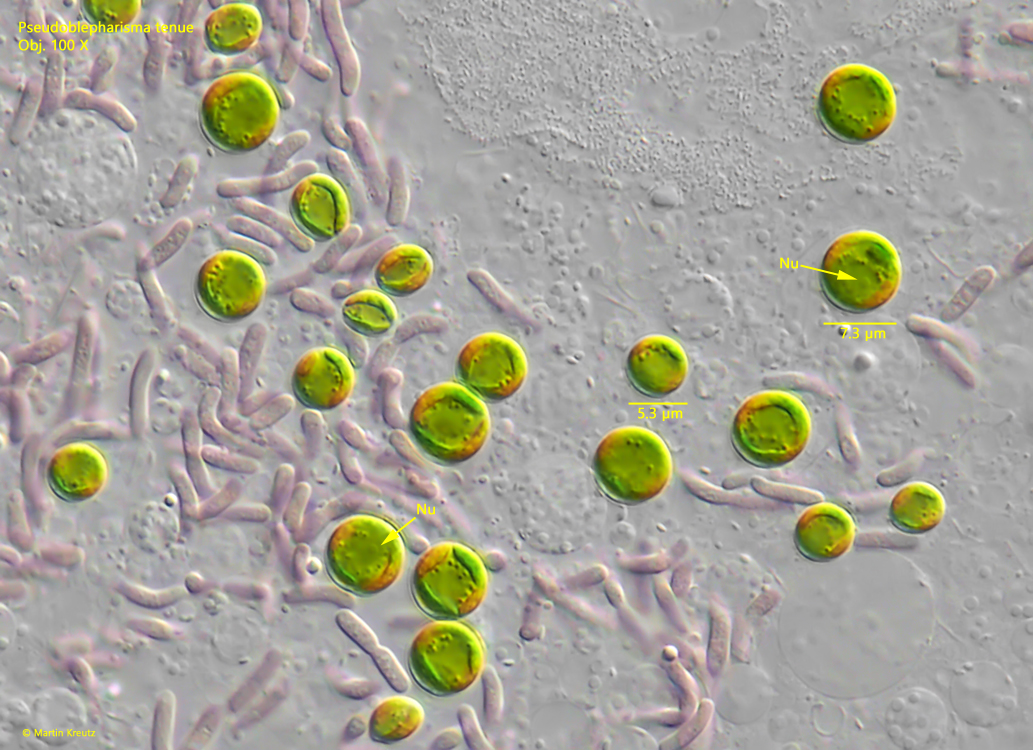
Fig. 6:Pseudoblepharisma tenue. The symbiotic algae are members of the genus Chlorella. In all cells a nucleus (Nu) is present The diameter of the algae is 5–7.5 µm. Obj. 100 X.
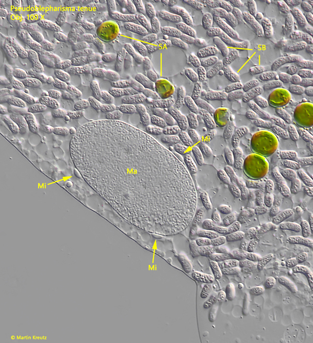
Fig. 7:Pseudoblepharisma tenue. The macronucleus (Ma) with three adjacent micronuclei (Mi). The micronuclei are lenticular. Obj. 100 X.
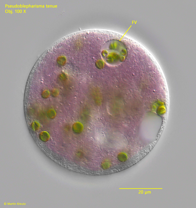
Fig. 8:Pseudoblepharisma tenue. In the cytoplasm several food vacuoles (FV) are visible filled with colorless bacteria. Obj. 100 X.
Within the population of Pseudoblepharisma tenue I found regularly dwarf forms, especially in old samples (s. figs. 9 a-b and 10 a-b). In most cases the dwarf forms had a length of 70–80 µm. The macronucleus in these specimens is enlarged and the adoral zone elongated or reduced. The reasons for the occurrence of the dwarf forms remained unclear to me. It is possible that they develop after many cell divisions, as it is known from other ciliates, in order to produce specimens of normal size again after conjugation. It is also possible that the dwarf forms develop due to unfavorable environmental conditions (e.g. lack of food, excessive oxygen content) and represent a preliminary stage for encystment.
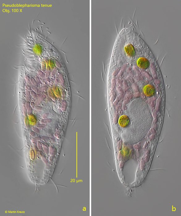
Fig. 9 a-b:Pseudoblepharisma tenue. L = 83 µm. A dwarf specimen with an adoral zone reaching to mid-body and a large macronucleus. Obj. 60 X.
Fig. 10 a-b:Pseudoblepharisma tenue. L = 70 µm. A second dwarf specimen with a reduced adoral zone and an enlarged macronucleus. Obj. 100 X.
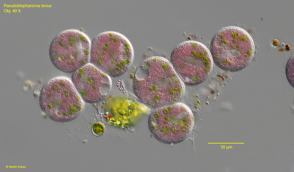
I found a lot of cysts of Pseudoblepharisma tenue in old samples (s. fig. 11). Their striking purple coloration makes them immediately noticeable and unmistakable. The diameter of the cysts is about 40–50 µm. The surface is smooth and without structure. Inside, in addition to the symbionts, the macronucleus and contractile vacuole are clearly visible. I examined many of these cysts and was able to find food vacuoles in some of them, which obviously contained the symbionts, i.e. either the bacteria (s. fig. 12 a-b) or the algae (s. fig. 13). It is possible that these are partially degraded and digested during the encystment process.
Fig. 11:Pseudoblepharisma tenue. An assemble of slightly squashed cysts. In the cysts the macronucleus and contractile vacuole as well as the symbiotic bacteria and algae are visible. Obj. 40 X.
Fig. 12 a-b:Pseudoblepharisma tenue. Two different cysts in detail. In the cysts food vacuoles (FV) are visible filled with symbiotic purple sulfur bacteria intended for digestion. Ma = macronucleus. Obj. 100 X.
Fig. 13:Pseudoblepharisma tenue. A further cyst with a food vacuole filled with symbiotic algae intended for digestion. Obj. 100 X.
Although I have seen thousands of specimens of Pseudoblepharisma tenue, I have only been able to find a single cell division (s. fig. 14 a-c). However, the specimen was already suffering and somewhat deformed. It is possible that Pseudoblepharisma tenue does not divide in the sample containers because anaerobic conditions are not present.
Fig. 14 a-c:Pseudoblepharisma tenue. A specimen during cell division. Obj. 100 X.
I was involved in the study of Pseudoblepharisma tenue by Muñoz-Gómez et al. (2021) and collected data on body length and the number of symbionts. To measure the body length and width, I photographed free-swimming specimens and later measured them on the photos. This minimizes measurement errors with moving objects:
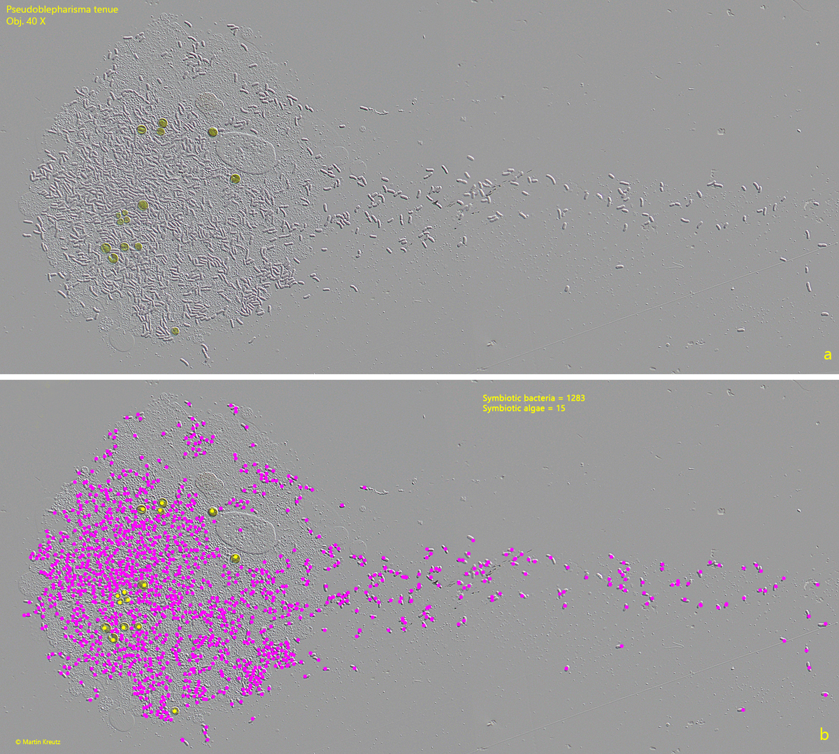
To count the symbionts, I squeezed the cells strongly to spread the bacteria and algae sufficiently. I took photos from each squashed specimen. Then, using a mouse click counter, I marked the symbiotic bacteria and algae in different colors with a dot so that they were not counted twice (s. fig. 15 a-b).
Fig. 15 a-b:Pseudoblepharisma tenue. For counting of the symbiotic bacteria and algae they were labelled with a pink or yellow dot to avoid double counting. Obj. 40 X.