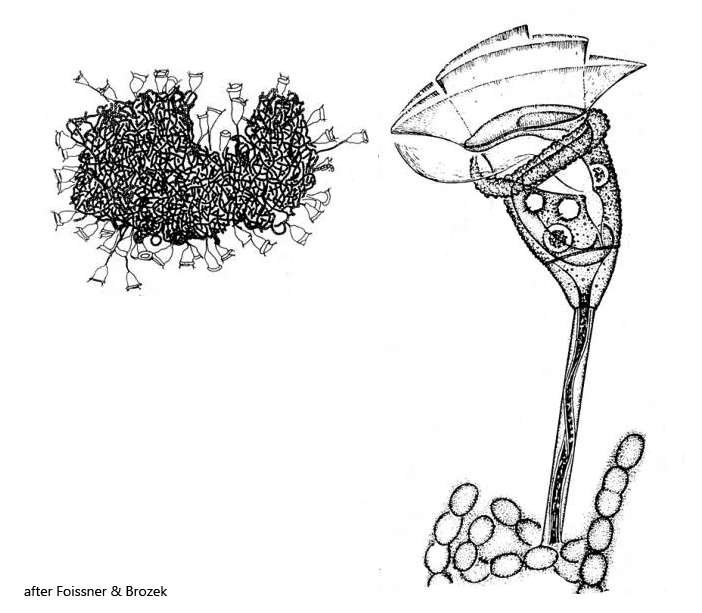
one micronucleus near anterior end of macronucleus
two contractile vacuoles
pellicle with conspicuous tubercles of various size
stalk up to 200 µm long, unbranched and smooth, contracts sinuously due to distinct myoneme
solitary, sessile on planktonic Anabaena coenobia
Pseudohaplocaulus infravacuolatus
Pseudohaplocaulus infravacuolatus is enormously common in Lake Constance and can be found in the plankton throughout the year. However, I have never found this peritriche ciliate in other localities. One finds the pseudocolonies exclusively on floating coenobia of the cyanobacterium Anabaena (likely Anabaena flos-aquae) on which the specimens have settled. Some Anabaena coenobia carry up to 50 specimens. The cells in the population from Lake Constance were on average 50–60 µm in size. However, some cells reached a length of 70 µm. The species can be identified partly by its planktonic life on Anabaena spec. but also by the irregularly sized blister-shaped pellicular tubercles with which the pellicle is covered (s. fig. 7 a-d and fig. 8). Also the two contractile vacuoles are characteristic and that the stalk does not (as in Vorticella) contract in spirals, but is S-shaped.
Fig. 1: Pseudohaplocaulus infravacuolatus. A pseudocolony on a freely floating Anabaena spec. coenobium. Obj. 20 X.
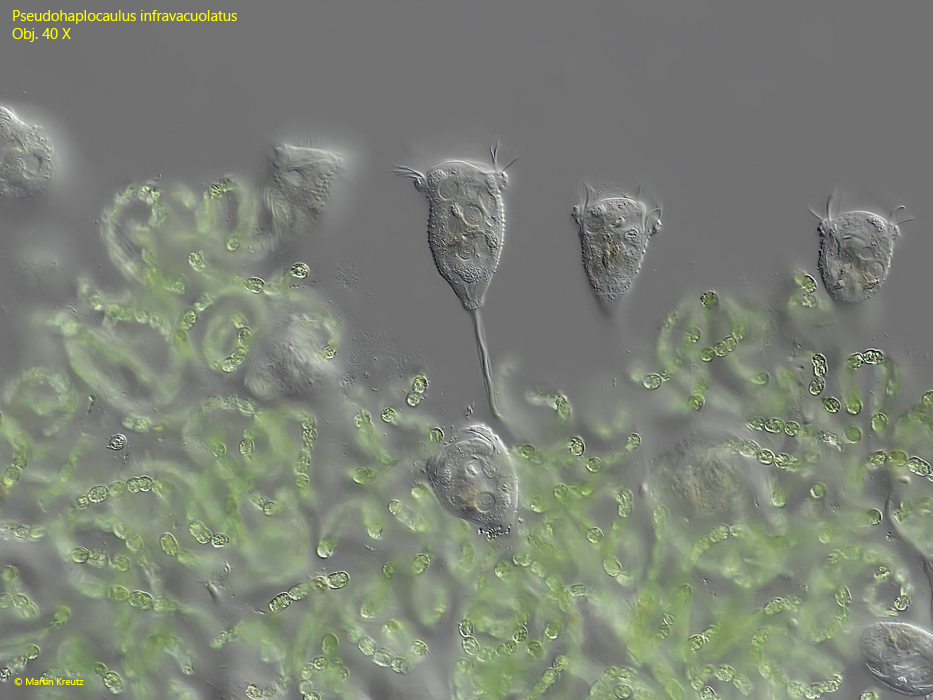
Fig. 2: Pseudohaplocaulus infravacuolatus. A pseudocolony on a slighty squashed Anabaena spec. coenobium. Obj. 40 X.
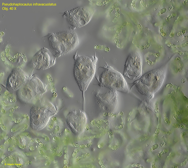
Fig. 3: Pseudohaplocaulus infravacuolatus. A second pseudocolony on a slightly squashed Anabaena spec. coenobium. Obj. 40 X.
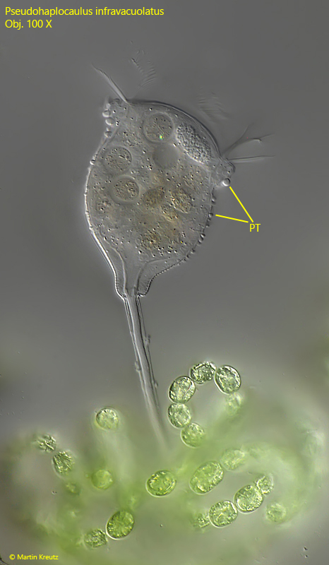
Fig. 4: Pseudohaplocaulus infravacuolatus. L = 55 µm. A fully extended specimen in detail. The pellicle is covered with blister-shaped pellicular tubercles (PT) of different sizes. Obj. 100 X.
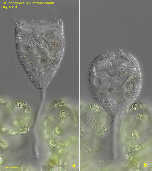
Fig. 5: Pseudohaplocaulus infravacuolatus. L = 56 µm. An extended (a) and contracted specimen (b) attached to an Anabaena spec. coenobium. Obj. 100 X.
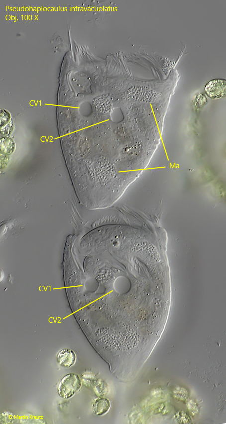
Fig. 6: Pseudohaplocaulus infravacuolatus. L = 52 µm. Two specimens with the clearly visible two contractile vacuoles (CV1, CV2) in each of them. Obj. 100 X.
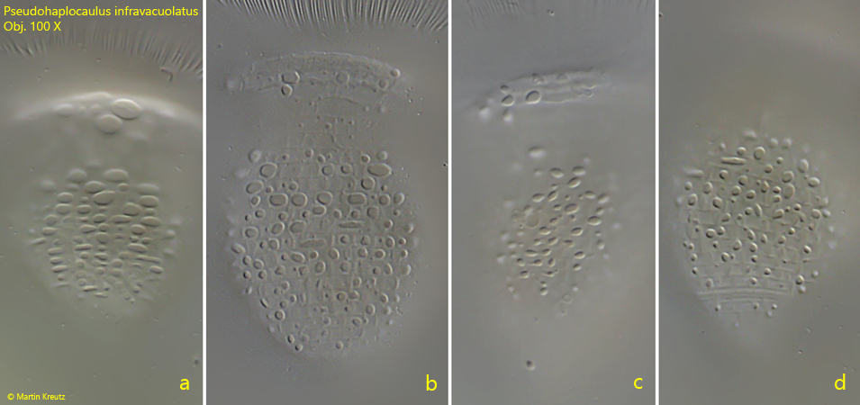
Fig. 7 a-d: Pseudohaplocaulus infravacuolatus. The different patterns of the pellicular tubercles of four specimens. Obj. 100 X.
Fig. 8: Pseudohaplocaulus infravacuolatus. The pellicular tubercles (PT) of a slightly squashed specimen in detail. Obj. 100 X.