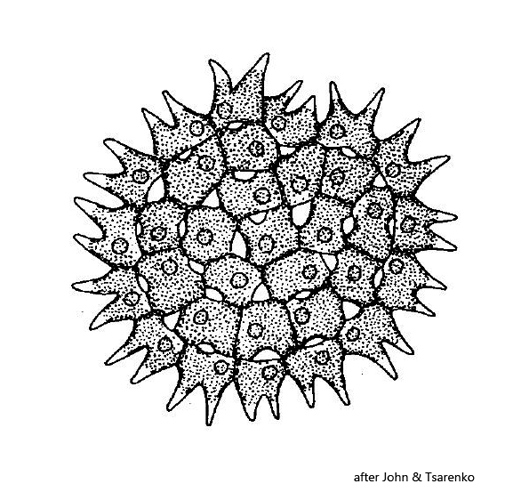
cells 7 – 23 X 7 – 22 µm, cell wall smooth or finely granulated
inner cells concentrically arranged
marginal cells bilobed
marginal cells occasionally bearing tufts of fine fibers at tips of lobes
intercellular space lens-shaped or triangular
chloroplast parietal
single pyrenoid
Pseudopediastrum cornutum
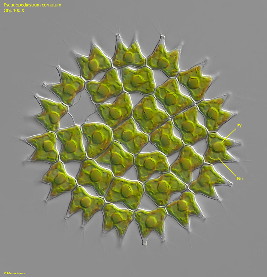
I found Pseudopediastrum cornutum in the plankton of the strongly eutrophic pond of the waste disposal company Constance. This pond is fed by the purified water of the sewage plant, which is still very rich in nutrients. Pseudopediastrum cornutum can be easily recognized by the H-shaped cells and the gaps between the cells, which are lens-shaped or triangular. The cells on the outer margin each bear two characteristic projections. In my population, these projections bore tufts of thin fibers, which I interpret as an adaptation to the planktonic lifestyle.
Fig. 1: Pseudopediastrum cornutum. D = 90 µm. A slightly squashed specimen. Nu = nucleus, PY = pyrenoid. Obj. 100 X.
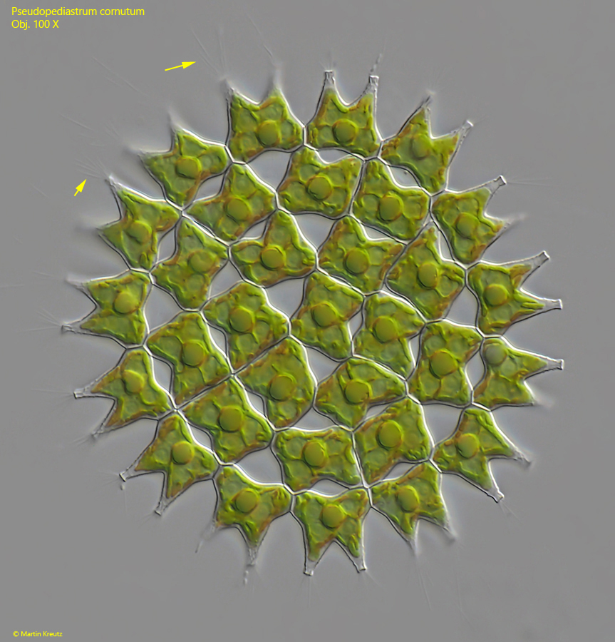
Fig. 2: Pseudopediastrum cornutum. D = 94 µm. A second slightly squashed specimen. Note the tufts of mucilaginous spines at the lobes of the marginal cells (arrows). Obj. 100 X.