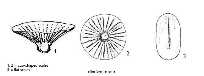
body covered with oval or elliptical, flat scales (1.2–2.0 x 0.7–1.1 µm)
cup-shaped scales (about 3.6 x 2.7 µm) arranged radially in a delicate mucilaginous sheath
numerous axopodia, about 25–30 µm long with granules
one contractile vacuole
one nucleus with a central nucleolus, excentrically located
cytoplasm contains often green or yellowish small algae
surface of protoplast covered with fine granules
Pseudoraphidocystis flabellata
I have found Pseudoraphidocystis flabellata only once in September 2020 in the Simmelried. It was several specimens which had settled on the floating coverslip.
This centrohelid heliozoan is very small. The protoplast of my specimens had a diameter of about 10 µm. In the unsquashed specimens one can see that a sheath of scales is present (s. fig. 1 a-b) but a precise examination and classification is only possible in squashed specimens (s. figs. 2 a-b and 3). Pseudoraphidocystis flabellata has two types of scales. The body is covered with flat, oval or ellipsoidal scales, which are hardly visible in the light microscope (s. fig. 2 a). The second kind of scales is cup-shaped (s. fig. 2 a-b). It can also be compared to the shape of a lampshade. These scales are arranged radially in a delicate mucilage layer, which, however, cannot be seen in DIC. Typical of these cup-shaped scales is a short, rounded stalk, which allows a clear assignment even with the light microscope.
The structure of the protoplast of Pseudoraphidocystis flabellata was previously unknown because the species was apparently described on the basis of electron microscopic analyses of the scales in fixed material. Therefore Siemensma (2021) writes “details of protoplast unknown” and gives only drawings of the scales (s. drawings above). However, in my live material I could recognize the structure of the protoplast well. The nucleus lies excentrically in the cell and has a central nucleolus (s. fig. 3). There is only one contractile vacuole, which lies peripherally. In the cytoplasm of all observed specimens I could find small phagocytozed algae. Only in strongly squashed specimens I could detect fine granules on the surface of the protoplast (s. fig. 3). The axopodia were about 25–30 µm long in the specimens of my population and regularly covered with granules (s. figs. 1 a-b and 2 a-b).
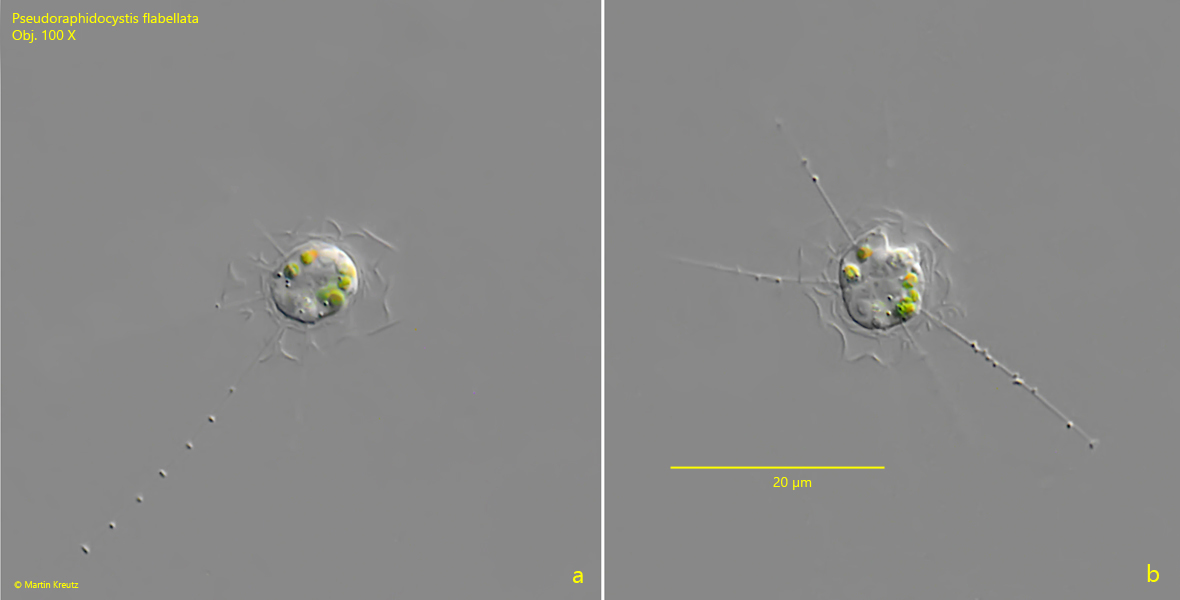
Fig. 1 a-b:Pseudoraphidocystis flabellata. D = 9.2 µm (of protoplast). Two focal planes of an unsphashed specimen. Obj. 100 X.
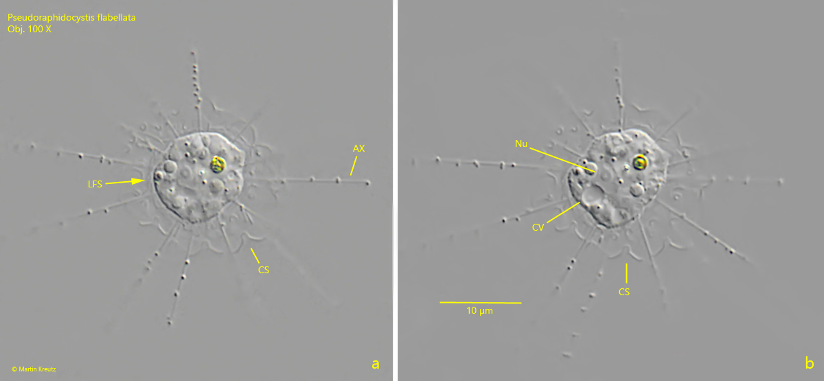
Fig. 2 a-b:Pseudoraphidocystis flabellata. D = 11.1 µm (of protoplast). Two focal planes of a slightly squashed specimen. Note the layer of flat scales (LFS) covering the protoplast and the radially arranged, cup-shaped scales (CS). AX = axopodia, CV = contractile vacuole, Nu = nucleus. Obj. 100 X.
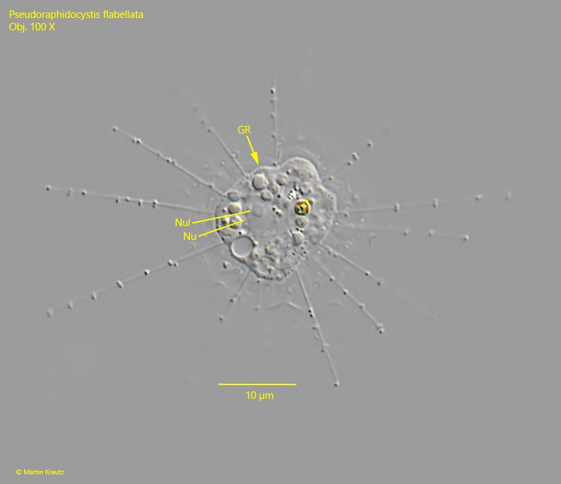
Fig. 3:Pseudoraphidocystis flabellata. In a squashed specimen the fines granules (GR) covering the protoplast are visible. Nu = nucleus, Nul = nucleolus. Obj. 100 X.