pellicular tubercles spherical, widely spaced, different sizes
solitary or in pseudocolonies
Pseudovorticella fasciculata
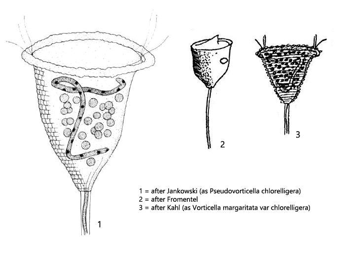
The species Pseudovorticella fasciculata has undergone many name changes and synonyms since its discovery by Müller in 1773. Müller himself placed the species in the to the genus Vorticella (Vorticella fasciculata). Later, Kahl stated that Vorticella fasciculata is apparently identical to the species Vorticella margaritata var. chlorelligera described by him.
Only in 1975 Foissner and Schiffmann assigned this species on the basis of the silver line system to the genus Pseudovorticella. The genus Pseudovorticella has a latticed silver line system, whereas species of the genus Vorticella possess a transverse stripe pattern. Subsequently, Jankowski (1976) renamed Vorticella margaritata var. chlorelligera to Pseudovorticella chlorelligera. Finally, in 1996 Foissner and Brozek re-introduced the original species name “fasciculata” which led to the current name Pseudovorticella fasciculata.
There are only few records and descriptions of Pseudovorticella fasciculata. I found this species in 1997 in the Simmelried. One could see a green fringe near the surface in the samples with the naked eye, where the pseudocolonies of Pseudovorticella fasciculata settled. They showed a clear positive phototactic reaction, because they always collected on the side of the vessel facing the light.
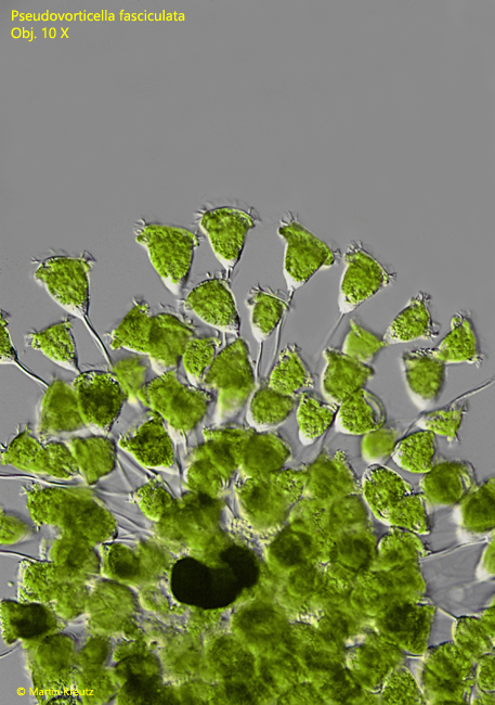
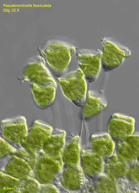
Fig. 1-2: Pseudovorticella fasciculata. L = 65–70 µm. Freely moving specimens of a pseudocolony. Obj. 10 X (fig. 1), Obj. 20 X (fig. 2).
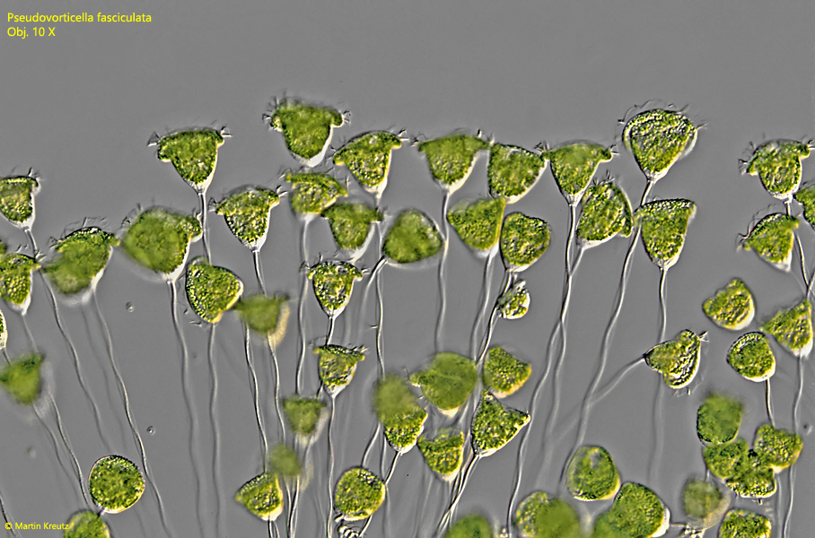
Fig. 3: Pseudovorticella fasciculata. L = 65–70 µm. Freely moving specimens of a second pseudocolony. Obj. 10 X.
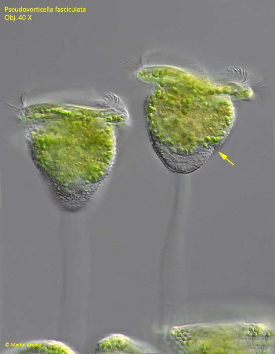
The cells in my population had a length of 60–70 µm. Kahl gives 60 µm for the synonymous species Vorticella margaritata var. chlorelligera and Jankowski gives up to 80 µm for the synonymous species Pseudovorticella chlorelligera. Due to the symbiotic algae the individuals show an intense green coloration. The body is broadly bell-shaped. A constriction in the posterior third of the body is conspicuous (s. fig. 4). Below this constriction the protoplasm is clear and free of symbiotic algae. In lateral view, this gives the impression of a crescent-shaped, clear plasma fringe. This is a very characteristic feature of Pseudovorticella fasciculata and has also been aptly drawn by Kahl in the synonymous species Vorticella margaritata var. chlorelligera (s. above, drawing 3, after Kahl).
Fig. 4: Pseudovorticella fasciculata. L = 63–67 µm. Freely moving specimens of a pseudocolony. Note the contriction at the posterior third of the cell (arrow) and the clear zone of cytoplasm free from symbiotic algae below this constriction. Obj. 40 X.
Fig. 5: Pseudovorticella fasciculata. Several contracted cells of a pseudocolony. The focal plane is on the pellicular tubercles. The blister-shaped tubercles are hard to see due to the green background. Obj. 40 X.
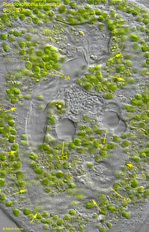
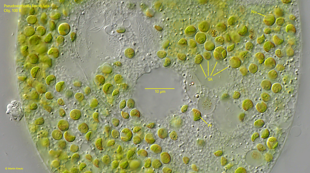
The number of contractile vacuoles has not been reported by previous authors. In the squeezed specimens I could clearly see two contractile vacuoles (s. fig. 6). The macronucleus is J-shaped. I could not identify a micronucleus. The symbiotic algae are spherical and very small with a diameter of 3–4 µm. They have a single chloroplast with one pyrenoid and a nucleus (s. fig. 7). The color of the symbiotic algae ist green yellowish.
Fig. 6: Pseudovorticella fasciculata. In a squashed cell the two contractile vacuoles are visible (CV 1, CV 2). The J-shaped macronucleus (Ma) is labelled by arrows. Obj. 100 X.
Fig. 7: Pseudovorticella fasciculata. The symbiotic algae (SA) in detail. The algae have a diameter of 3–4 µm and have a single chloroplast with a pyrenoid (PY) and a nucleus (Nu). The color of them is green yellowish. Obj 100 X.