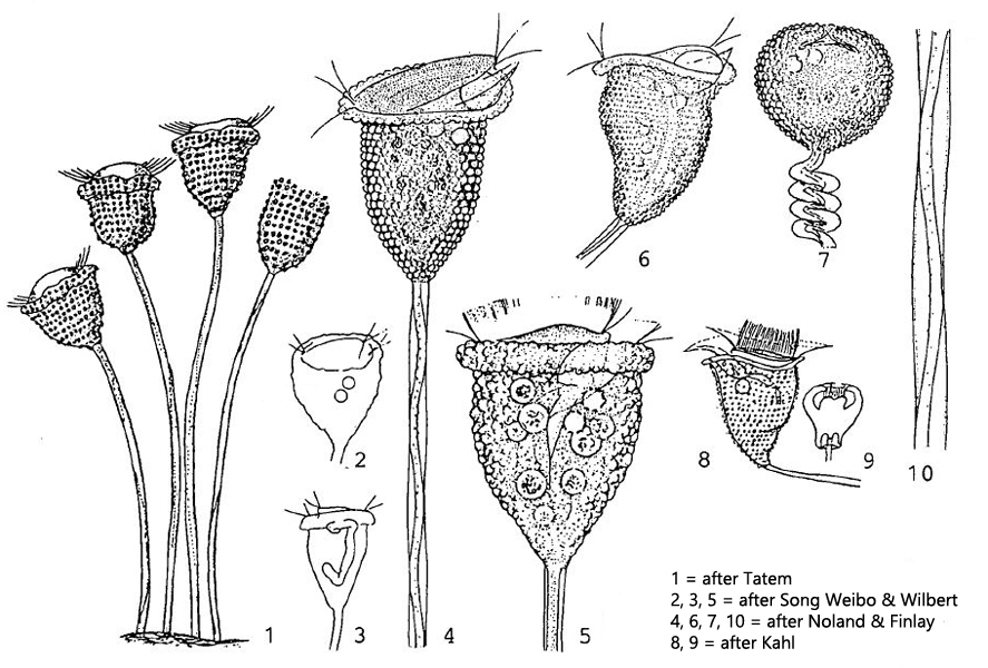
pellicle covered with blister-shaped pellicular tubercles
tubercles of different size, often containing an oily droplet
spirally contractile stalk
forming pseudocolonies
Pseudovorticella monilata
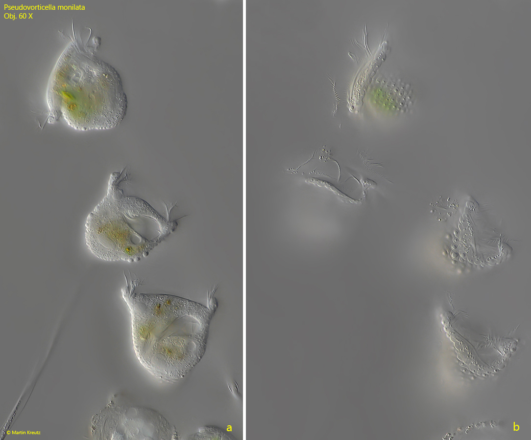
I found pseudocolonies of Pseudovorticella monilata 2022 in old samples from Simmelried. These pseudocolonies of 50–100 zooids were found on small detritus flakes on the vessel wall as well as on the surface. At low magnifications, Pseudovorticella can be confused with Vorticella, due to the spirally contracting stalk. Only at higher magnifications (about Obj. 40 X) do the vesicular pellicular tubercles covering the surface of the zooids become visible. These pellicular tubercles form the major distinguishing feature between the genera Vorticella and Pseudovorticella. The pellicle of species of the genus Vorticella is usually finely striate, without vesicular tubercles. Finally, the species Pseudovorticella monilata is distinguished by the presence of oil-like droplets in the pellicular tubercles, the presence of two contractile vacuoles and the J-shaped macronucleus.
Fig. 1 a-b: Pseudovorticella monilata. L = 53–61 µm. Freely moving specimens of a pseudocolony in two focal planes. Obj. 60 X.
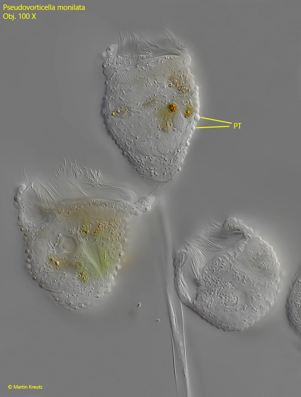
Fig. 2: Pseudovorticella monilata. L = 56 µm. Freely moving specimens of a pseudocolony. In lateral view the blister-shaped pellicular tubercles (PT) are visible, covering the zooids. Obj. 100 X.
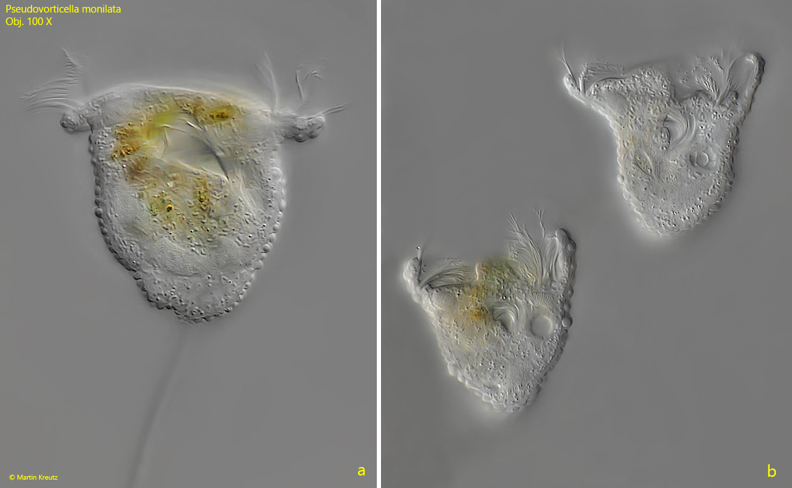
Fig. 3 a-b: Pseudovorticella monilata. L = 52–61 µm. Freely moving zooids in detail. Obj. 100 X.
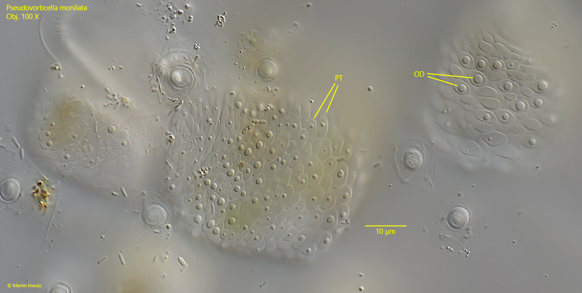
Fig. 4: Pseudovorticella monilata. The blister-shaped pellicular tubercles (PT) in a squashed specimen. The tubercles often contain a drop of an oily liquid (OD) believed to be paraglycogen. Obj. 100 X.
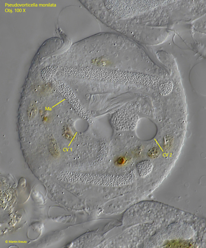
Fig. 5: Pseudovorticella monilata. In this strongly squashed specimen the two contractile vacuoles are visible (CV 1, CV 2) and the J-shaped macronucleus (Ma). Obj. 100 X.