Raphidiophrys elegans

(Hertwig & Lesser, 1874) emend. Penard, 1904

Most likely ID: n.a.


Synonym: n.a.


Sampling location: Simmelried 


Phylogenetic tree: Raphidiophrys elegans


Diagnosis: 

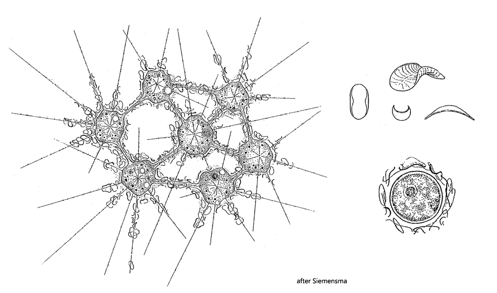
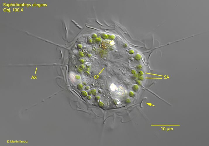
  • cells about 30 µm in diameter (without coat of scales)

  • scales 6.2–8.6 µm x 4.4–6.5 µm, broadly oval, sometimes oblong

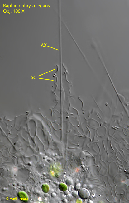
  • scales curved with the poles bent downwards, edges strongly inflected

  • scales covering the bases of the axopods

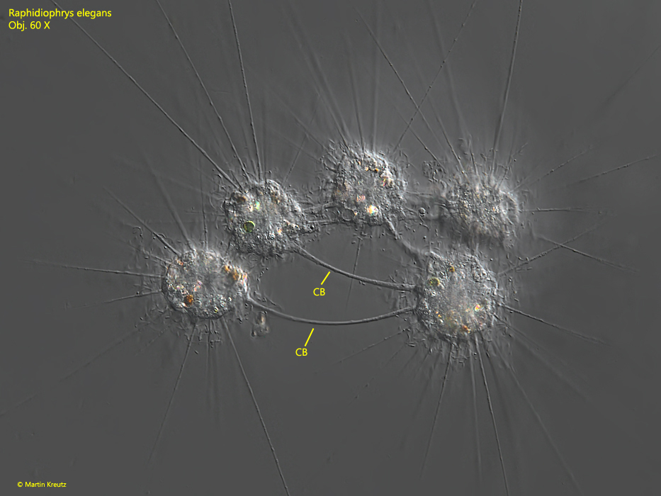
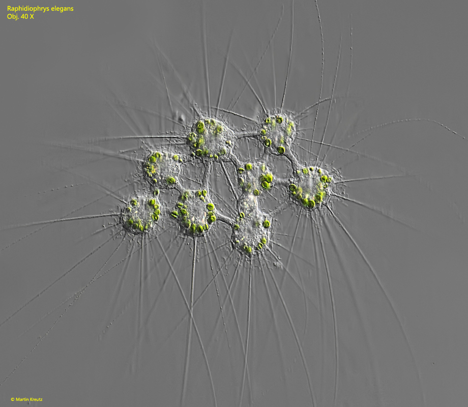
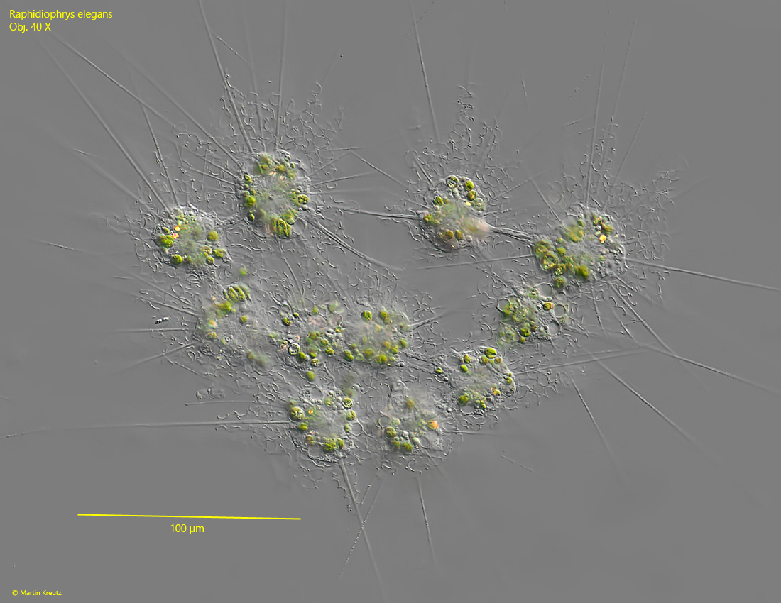
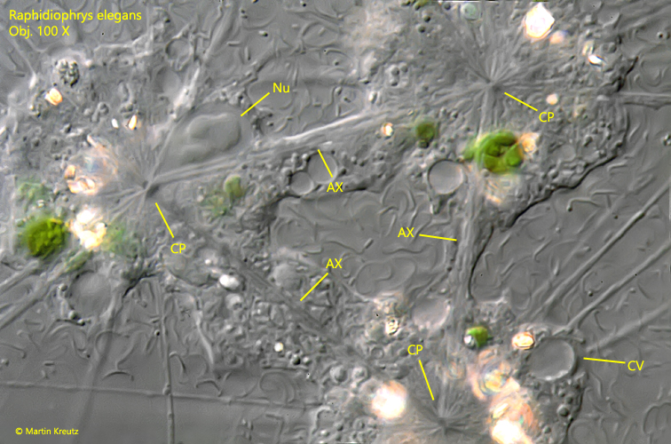
  • axopods up to 170 µm long

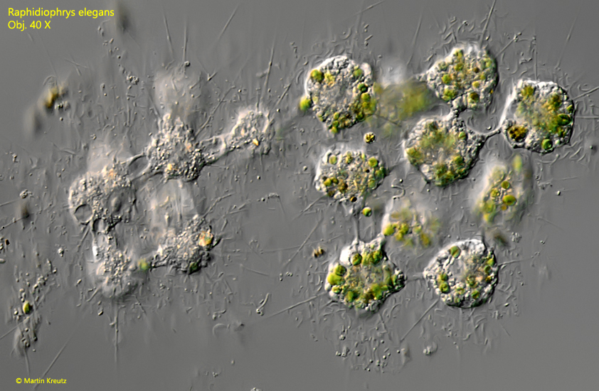
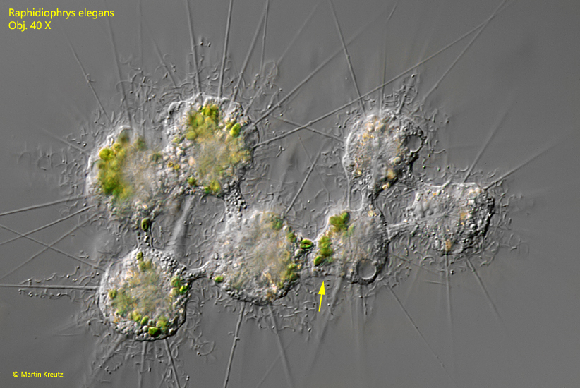
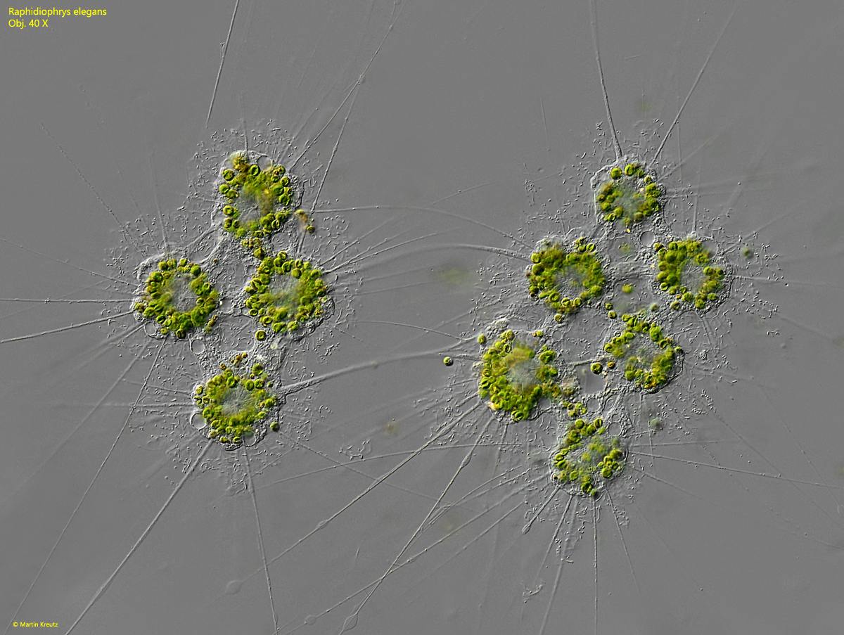
  • solitary as well as in colonies of up to 30 individuals connected via cytoplasmic bridges

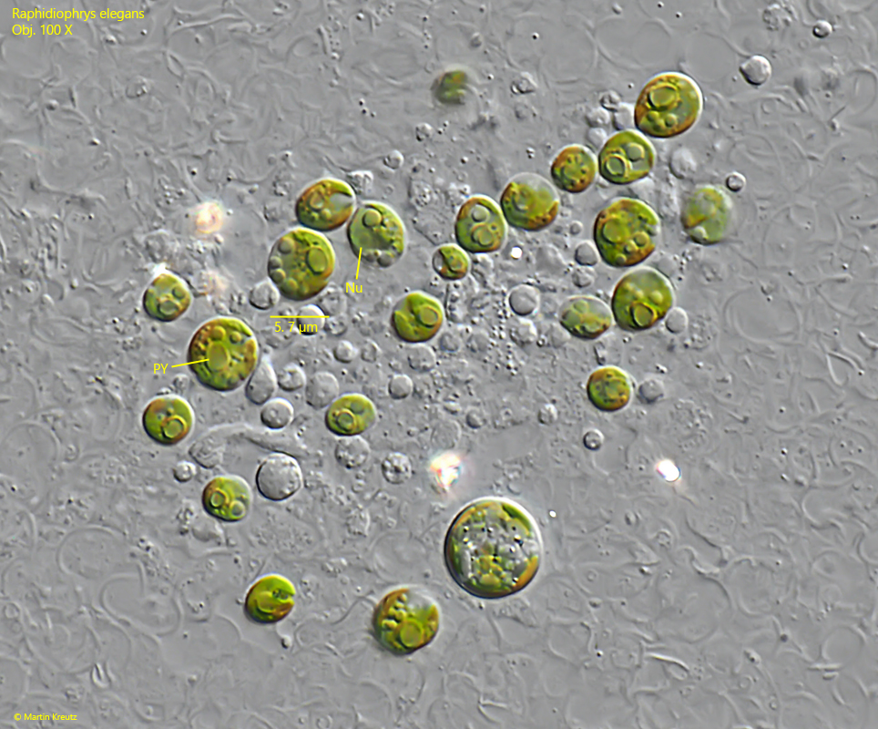
  • cells sometimes with symbiotic algae

  • centroplast in the center of the cell

  • nucleus in eccentric position

  • up to 5 contractile vacuoles

Raphidiophrys-elegans
Raphidiophrys elegans

I find colonies of Raphidiophrys elegans quite often, especially in old samples with plant material. After pipetting and transfer of the colonies on a slide it takes about 30–60 min. under high layer thickness before they are reorganized. Then an observation of the undisturbed colonies is possible. I find colonies with individuals lacking symbiotic algae (s. fig. 1) but also colonies with individuals containing symbiontic algae (s. figs. 2 and 7). Interestingly, there are also colonies containing both variants (s. fig. 5 and 6). In these colonies I could observe that the symbiotic algae can obviously be exchanged via the bridges between the cells (s. fig. 6). Why there are colonies without symbiotic algae remains to be investigated. Mostly I have found small colonies of less than 10 individuals and most of them also had symbiont algae. Food vacuoles can also be observed in the cytoplasmic bridges between the cells. Therefore, it can be assumed that phagocytized food is distributed among the cells. However, I could never observe prey capture or phagocytosis of already captured prey.


More images and information about Raphidiophrys elegans: Ferry Siemensma – Microworld – Raphidiophrys elegans

Raphidiophrys-elegans

Fig. 1: Raphidiophrys elegans. A colony of 4 cells without symbiotic algae connected via cytoplasmic bridges (CB). The individual cells have a diameter of 30–34 µm (without coat of scales). Obj. 60 X.

Raphidiophrys-elegans

Fig. 2: Raphidiophrys elegans. A colony of 8 cells with symbiotic algae connected via cytoplasmic bridges. The individual cells have a diameter of 32–36 µm (without coat of scales). Obj. 40 X.

Raphidiophrys-elegans

Fig. 3: Raphidiophrys elegans. A second colony of 10 cells with symbiotic algae connected via cytoplasmic bridges. The individual cells have a diameter of 34–40 µm (without coat of scales). Obj. 40 X.

Raphidiophrys-elegans

Fig. 4: Raphidiophrys elegans. A strongly squashed solitary specimen. In the center of the cell the centroplast (CP) ist visible organizing the microtubules for formation of the axpodia (AX). The scales have the shape of a “U” when optically sectioned along the longitudinal axis (arrow). SA = symbiotic algae. Obj. 100 X.

Raphidiophrys-elegans

Fig. 5: Raphidiophrys elegans. A colony which consists partly of individuals without and with symbiotic algae. Because of this composition, it seems likely that the colonies cannot only divide, but also merge. Obj. 40 X.

Raphidiophrys-elegans

Fig. 6: Raphidiophrys elegans. Another colony which consists of individuals with and without symbiotic algae. Obviously the cytoplasmic bridges are also used for the exchange of symbiotic algae (arrow). Obj. 40 X.

Raphidiophrys-elegans

Fig. 7: Raphidiophrys elegans. Two colonies connected via long cytoplasmic bridges. It remains unclear if this colony is a dividing or if two colonies are merging.  Obj. 40 X.

Raphidiophrys-elegans

Fig. 8: Raphidiophrys elegans. The bases of the axopodia (AX) are covered by a layer of scales (SC). Obj. 100 X.

Raphidiophrys-elegans

Fig. 9: Raphidiophrys elegans. The broadly oval scales in a strongly squashed specimen. Obj. 100 X.

Raphidiophrys-elegans

Fig. 10: Raphidiophrys elegans. The symbiotica agla contains a single pyrenoid (PY) and a nucleus (Nu). The shape of the symbiotic algae is not spherical but slightly irregular. Obj. 100 X.

Raphidiophrys-elegans

Fig. 11: Raphidiophrys elegans. The structure of the cytoplasmic bridges in detail. The cells are connected via axopodia (AX), which originate in the centroplasts (CP) of the cells. The bridge of cytoplasm is then built along these axopodia, which are composed of microtubules. CV = contractile vacuole, Nu = nucleus. Obj. 100 X.