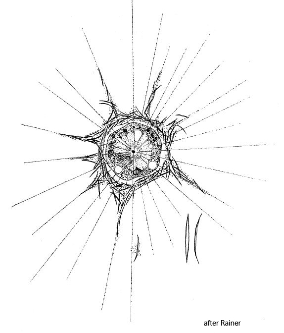
scales hollow, tapered to the ends, centrally with a 1.2 um wide suture
nucleus in eccentric position
centroplast in the center of cell
> 4 contractile vacuoles
Raphidocystis pallida
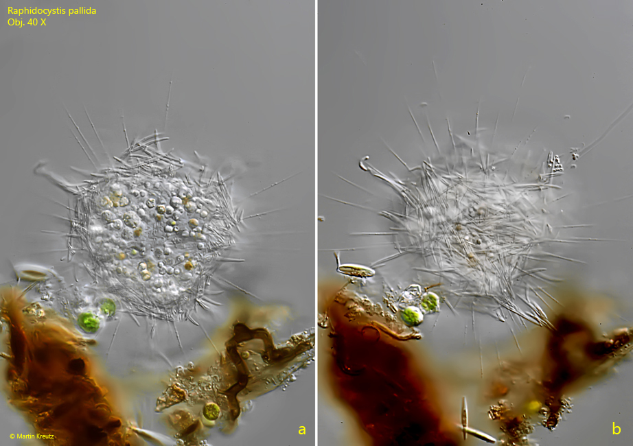
Raphidocystis pallida is described as a very common heliozoan, but I could find this species only a few times. I documented a finding in July 2000 in Ulmisried (s. fig. 1 a-c) and my last finding in September 2019 in the Mühlweiher Litzelstetten (s. figs. 2-5). Raphidocystis pallida is very easy to identify by the large, coarse scales, which are spindle-shaped and surround the cell in a thick layer. The scales are shaped like a rolled up sheet of paper. A suture is visible in the center, which looks like a slit in DIC (s. fig. 5). The species belongs to the centroheliozoa and therefore has a centroplast (s. fig. 4), which organizes the structure of the axopodia from microtubules.
Fig. 1 a-b:Raphidocystis pallida. D = 98 µm (with layer of scales). Two focal planes of a specimen attached to detritus. Obj. 40 X.
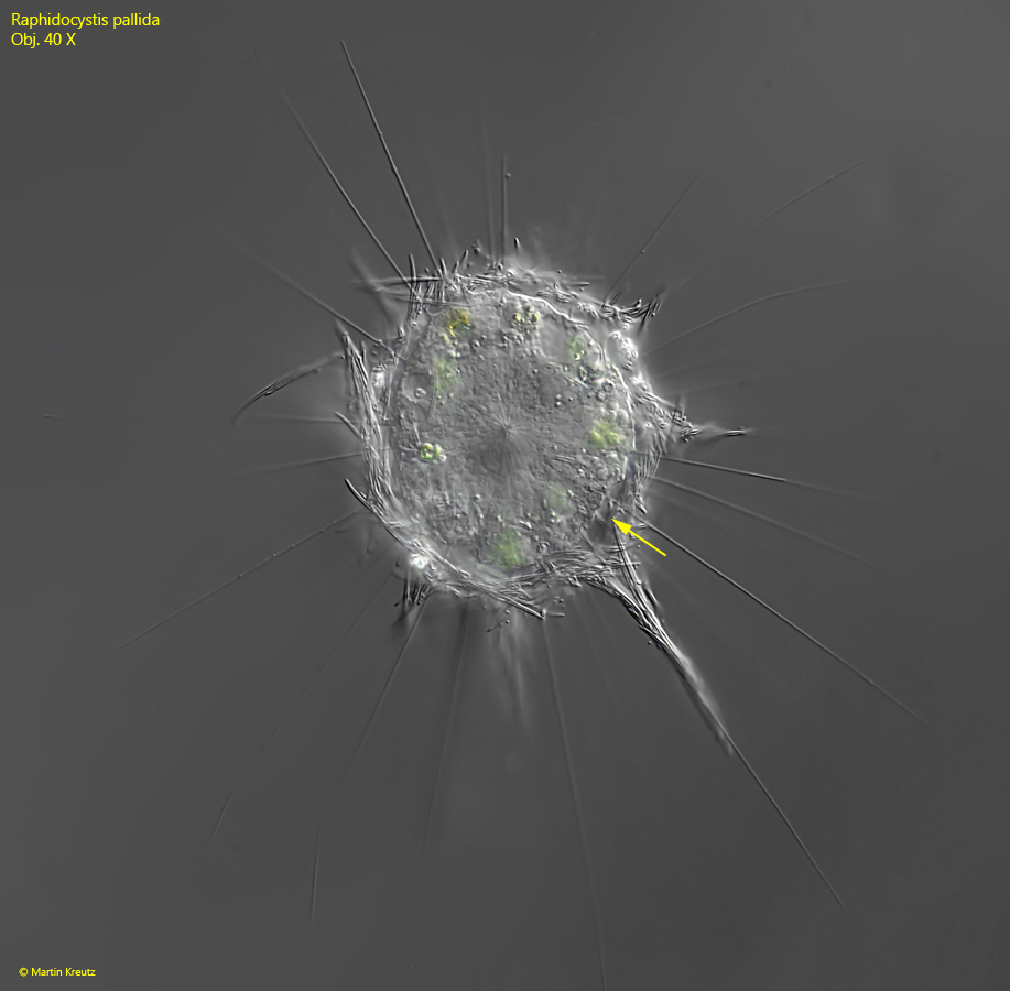
Fig. 2:Raphidocystis pallida. D = 105 µm (with layer of scales). A second specimen with focal plane on the center of the cell. Note the small gap between the cytoplasm of the cell and the layer of scales (arrow). Obj. 40 X.
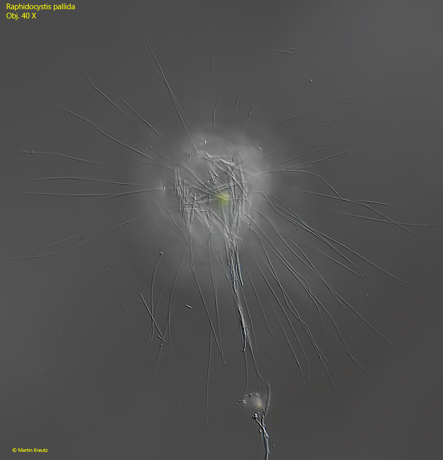
Fig. 3:Raphidocystis pallida. D = 105 µm (with layer of scales). The same specimen shown in fig. 2 with the focal plane on the extended axopodia. Obj. 40 X.
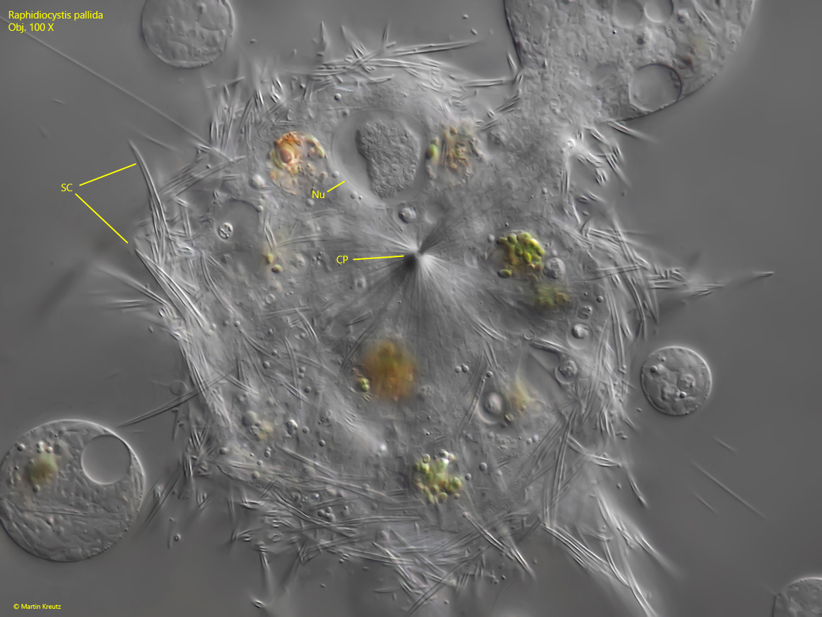
Fig. 4:Raphidocystis pallida. A strongly squashed specimen. CP = centroplast, Nu = nucleus in eccentric position, SC = layer of scales. Obj. 100 X.
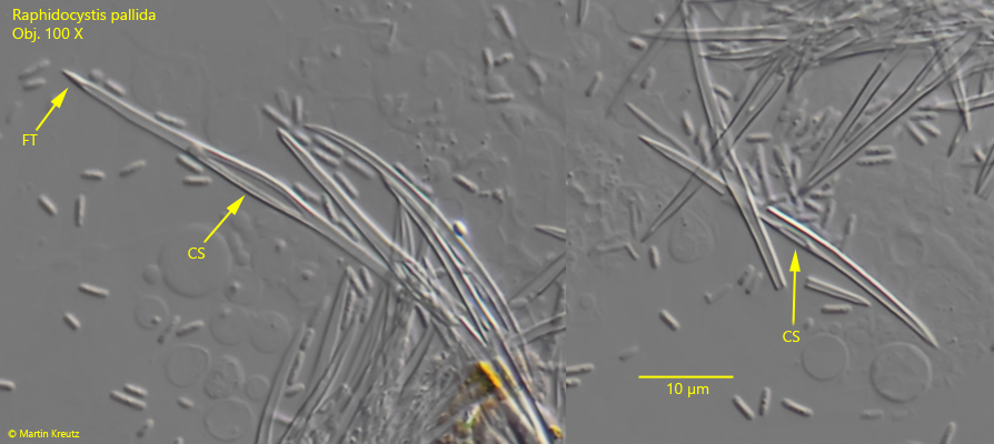
Fig. 5:Raphidocystis pallida. The scales in detail. In the center of the scales a slit-shaped suture is visible (CS). The scales are hollow, but the tips are filled (FT). Obj. 100 X.