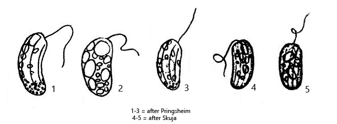
anterior end rounded or obliquely truncate, posterior end rounded
Ventral side weakly concave, dorsal side curved
length 13–25 µm
one flagellum, about body length
furrows of the pellicle widely spaced, only slightly twisted
several paramylon grains, often 2–3 larger grains
nucleus at posterior end
Rhabdomonas incurva
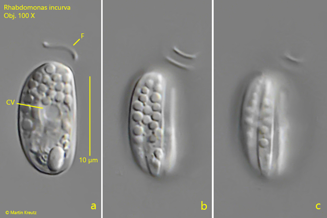
I have found Rhabdomonas incurva only rarely in the Simmelried. Rhabdomonas incurva belongs to the euglenoids and is easily recognized by its slightly bean-shaped appearance. The furrows on the pellicle are sometimes difficult to see when the cell is filled with many paramylon grains. Rhabdomonas differs from the genus Menoidium in the shape of the cell in cross-section. While the cells of Rhabdomonas are almost circular in cross-section, members of the genus Menoidium are strongly laterally flattened.
Fig. 1 a-c:Rhabdomonas incurva. L = 14 µm. Three focal planes of a freely swimming specimen. Note the slightly twisted furrows of the pellicle (b, c). CV = contractile vacuole, F = flagellum. Obj. 100 X.
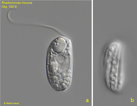
Fig. 2 a-b:Rhabdomonas incurva. L = 15 µm. Two focal planes of an almost straight specimen. Obj. 100 X.
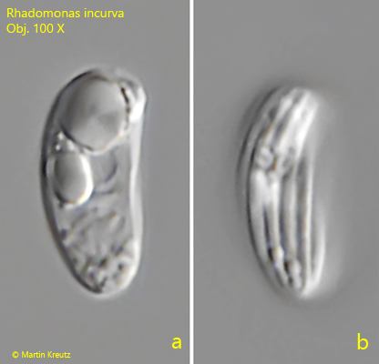
Fig. 3 a-b:Rhabdomonas incurva. L = 12 µm. A specimen with tighter arranged furrows of the pellicle. Obj. 100 X.