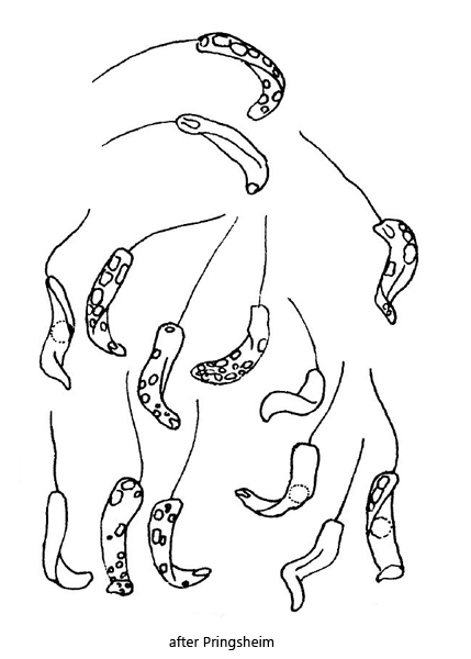
anterior end transversely blunted or broadly rounded
posterior end slightly tapered and pointed
one flagellum of body length
paramylon grains ovoid or short cylindrical
Rhabdomonas spiralis
So far I have found Rhabdomonas spiralis only in the Simmelried. However, the species occurs here only in large, temporal intervals, sometimes with several years in between. Because of the spiral shape and small size the species is easy to identify. Obviously only the original description by Pringsheim is available. In this he describes the difficulty to determine the exact shape of the cells and to recognize their internal structure.
It is noted by Huber-Pestalozzi (1955) that Rhabdomonas spiralis would have been better placed in the genus Menoidium because of the curved shape and the slight flattening of the cells. However, to my knowledge, this has not yet been done.
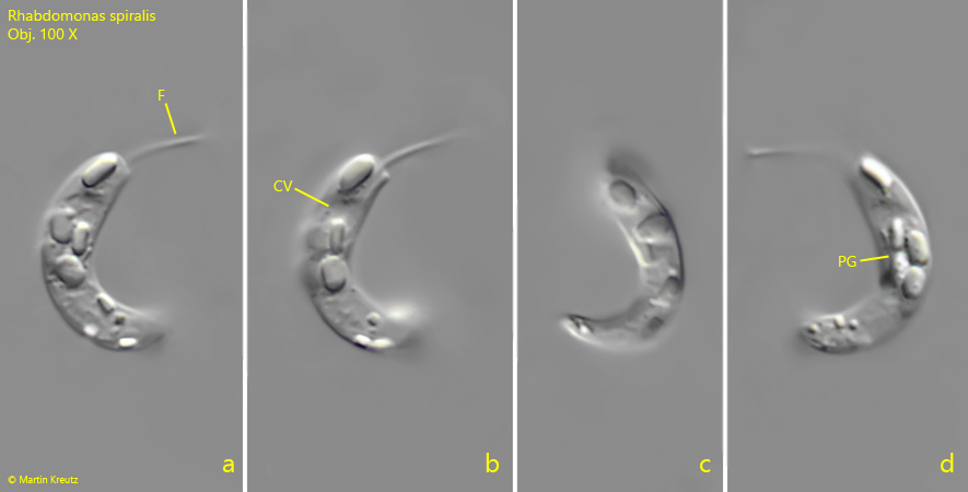
Fig. 1 a-d:Rhabdomonas spiralis. L = 15 µm. A freely swimming specimen. CV = contractile vacuole, F = flagellum, PG = paramylon grains. Obj. 100 X.
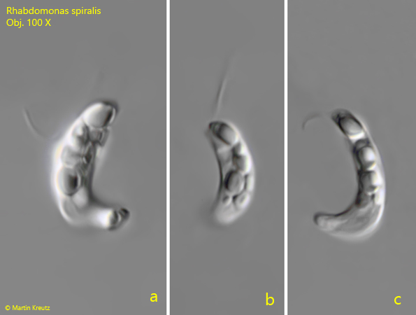
Fig. 2 a-c:Rhabdomonas spiralis. L = 16 µm. A second, freely swimming specimen. Obj. 100 X.
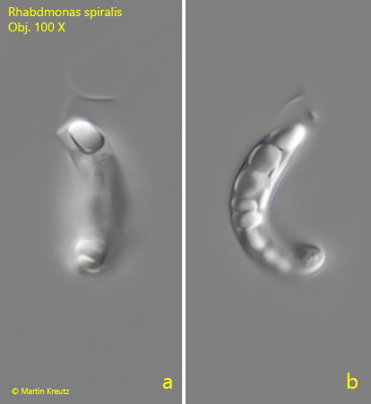
Fig. 3 a-b:Rhabdomonas spiralis. L = 15 µm. A third, freely swimming specimen. Obj. 100 X.