apically two distinct palps with acute distal ends, about 10–12 µm long
tight rows of somatic cilia
extrusomes absent
terminal cilia slightly elongated
macronucleus elongated ellipsoid, one adjacent micronucleus
contractile vacuole terminal
Rhinothrix barbatula
Rhinothrix barbatula is a very rare ciliate. I have only found 3 specimens in May 2003, July 2005 and November 2021. I found all three specimens in the Simmelried between decomposing plant masses.
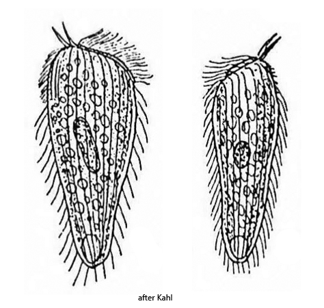
Apart from the specimens of Rhinothrix barbatula I found, there are very few records. The first description comes from Penard (1922), who only found one specimen and named the species Holophrya barbatula. His specimen had the typical apical palps, but no oral bulge was visible. Foissner et al. (2005) therefore assume that Penard found Holophrya sulcata or Holophrya pogonias, which also have palps. Kahl (1935) then found 3 further specimens on which he was able to demonstrate the inconspicuous oral bulge (s. drawings above) and therefore transferred the species to the genus Spathidium. Later, Spathidium barbatula was transferred to the genus Rhinothrix by Foissner et al. (2005), in which spathidiid ciliates with a palpe-like extension of the oral bulge are grouped together.
Rhinothrix barbatula is difficult to observe in vivo. The ciliate swims quickly and after the coverslip is applied, the two antenna-shaped palps are quickly melted. That means there is little time to photograph the free-swimming specimens before they denature. Squashed specimens also melt very easily, which makes precise examination at high magnifications more difficult. As a result, my yield of usable photos is also quite low.
In the first specimen found in May 2003 (s. figs. 1 a-b and 2), I was unable to detect the oral bulge. This led Foissner to assume that the taxonomic position is still uncertain, as it may not be a spathidiid ciliate after all. Only with the second specimen from July 2005 did I obtain better photos of the free-swimming specimen on which the two palps are clearly visible (s. fig. 3 a-e) and also evidence of the oral bulge, which is inconspicuous and flat (s. fig. 4). In the free-swimming specimen the oral bulge is not visible.
The three specimens from my population essentially correspond to the description by Kahl. The specimens were 108–122 µm long. The two palps were 10–12 µm long. Both have the same length. The macronucleus is elongated ellipsoid and about 30–35 µm long. The micronucleus, which is adjacent to the macronucleus, could only be found in the third specimen (s. fig. 7). All specimens were club-shaped and tapered in the posterior third. Kahl could not detect any extrusomes in his specimens. In my specimens I could detect rod-shaped extrusomes with a length of 7.8–8.4 µm in the first two specimens (s. figs. 2 and 5), but none in the third specimen. It is therefore unclear whether the extrusomes in the first two specimens possibly originate from prey organisms.
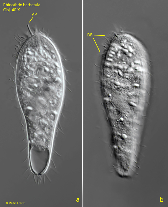
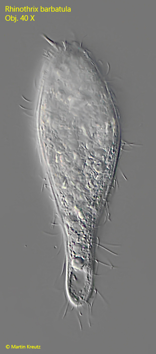
Fig. 1 a-b:Rhinothrix barbatula. L = 122 µm. A freely swimming specimen found in May 2003. Only one of the two apical palps (AP) is in the focal plane. DB = dorsal brush. Obj. 40 X.
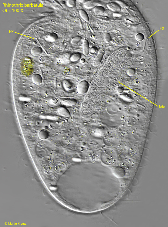
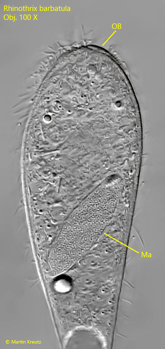
Fig. 2:Rhinothrix barbatula. The squashed specimen as shown in fig. 1 a-b. The macronucleus (Ma) is elongated ellipsoid and some rod-shaped extrusomes (EX) scattered in the cytoplasm are visible. Obj. 100 X.
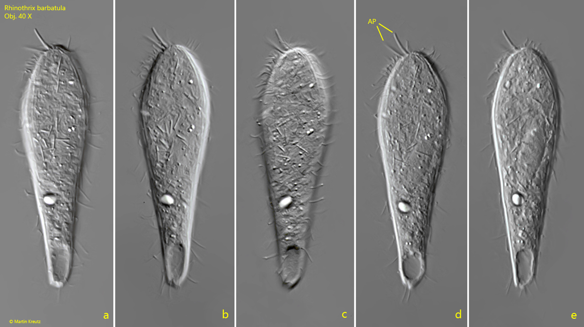
Fig. 3 a-e:Rhinothrix barbatula. L = 113 µm. The freely swimming second specimen found in July 2005. Note the both apical palps (AP) with a length of 11.8 µm. Obj. 40 X.
Fig. 4:Rhinothrix barbatula. Focal plane on the macronucleus (Ma) in the squashed specimen as shown in fig. 3 a-e. Apically the inconspicuous oral bulge is visible. Obj. 100 X.
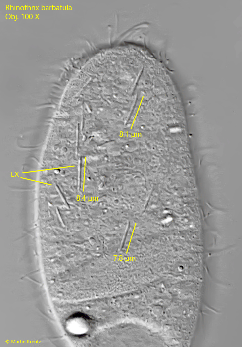
Fig. 5:Rhinothrix barbatula. Focal plane on the rod-shaped extrusomes (EX) in the specimen as shown in fig. 3 a-e. The extrusomes are 7.8 -8.4 µm long and have tapered ends. Obj. 100 X.
Fig. 6:Rhinothrix barbatula. L = 108 µm. The freely swimming third specimen found in November 2021. Obj. 40 X.
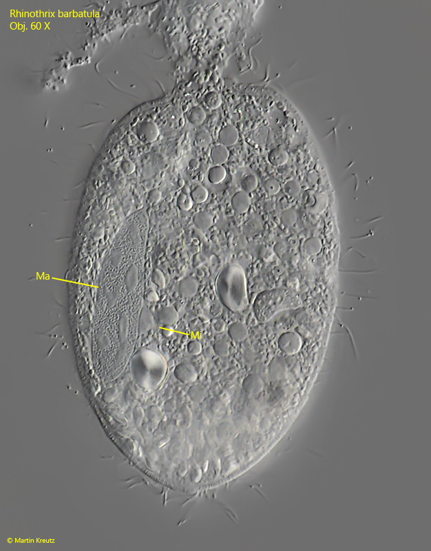
Fig. 7:Rhinothrix barbatula. Focal plane on the macronucleus (Ma) and micronucleus (Mi) in the squashed specimen as shown in fig. 6. In this specimen no extrusomes are visible. Obj. 100 X.