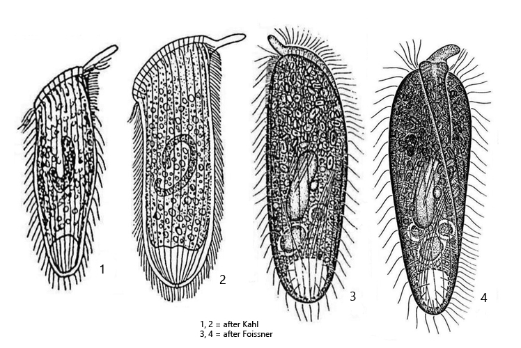
oral bulge with a distinct palp at dorsal side (length 5–12 µm)
ventrally the oral bulge running sigmoidal to right side of posterior end
accumulation of refractive inclusions in anterior third
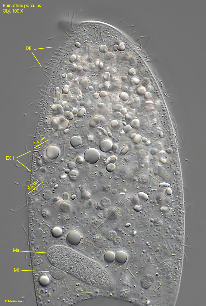
three types of extrusomes (type 1 = curved, about 7 µm, type 2 = slightly curved, dabout 50 µm, type 3 = straight, about 1.5 µm)
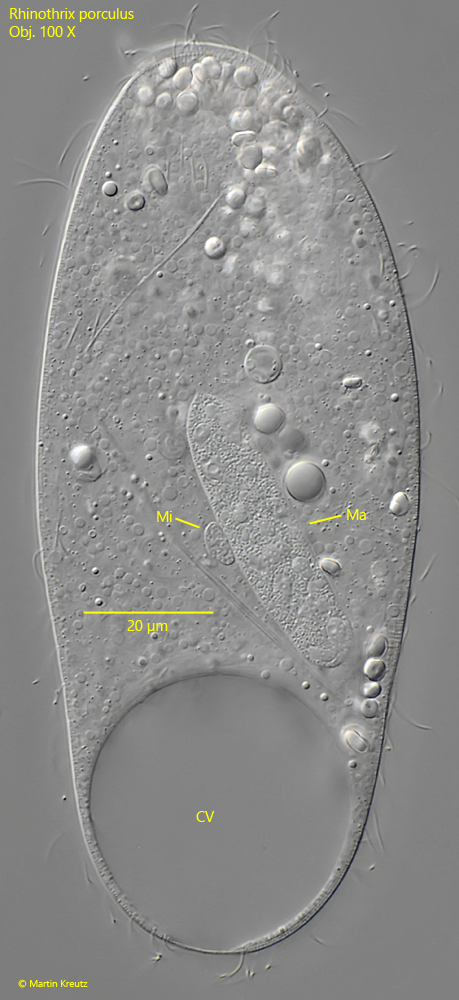
macronucleus oblong, rarely globular or reniform
one micronucleus almost hemi-spherical, adjacent to macronucleus
contractile vacuole terminal
Rhinothrix porculus
I find Rhinothrix porculus rarely, but regularly. All the specimens I have found so far come from the Simmelried. There I find Rhinothrix porculus in the uppermost mud layer and sometimes also between decomposing plant masses. I have not yet been able to find this ciliate in my other locations.
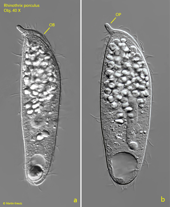
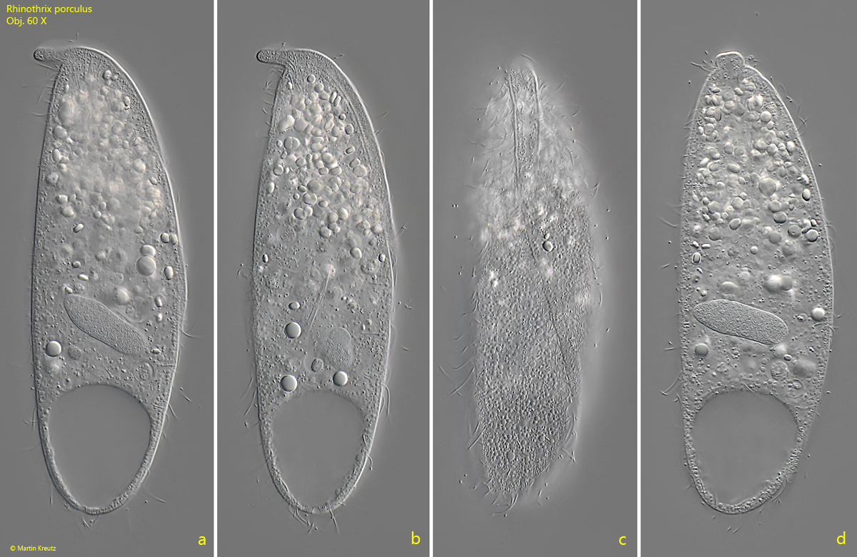
Rhinothrix porculus is easy to identify by its oral palp even at small magnifications (s. figs. 1 a-b, 2 a-d and 3 a-d). Confusion with other species is practically impossible. The oral palp is a dorsal extension of the oral bulge. This can also be recognized by the fact that it contains extrusomes of type 3 (s below). The ciliate swims quite slowly and rotates around its own axis.
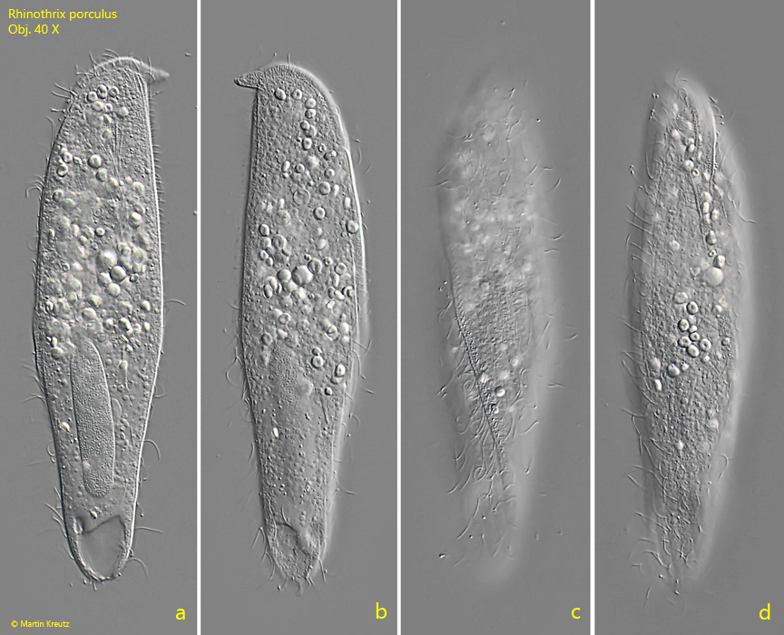
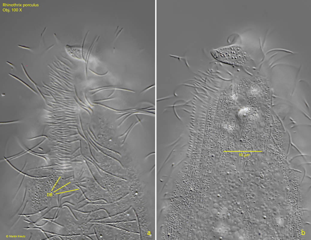
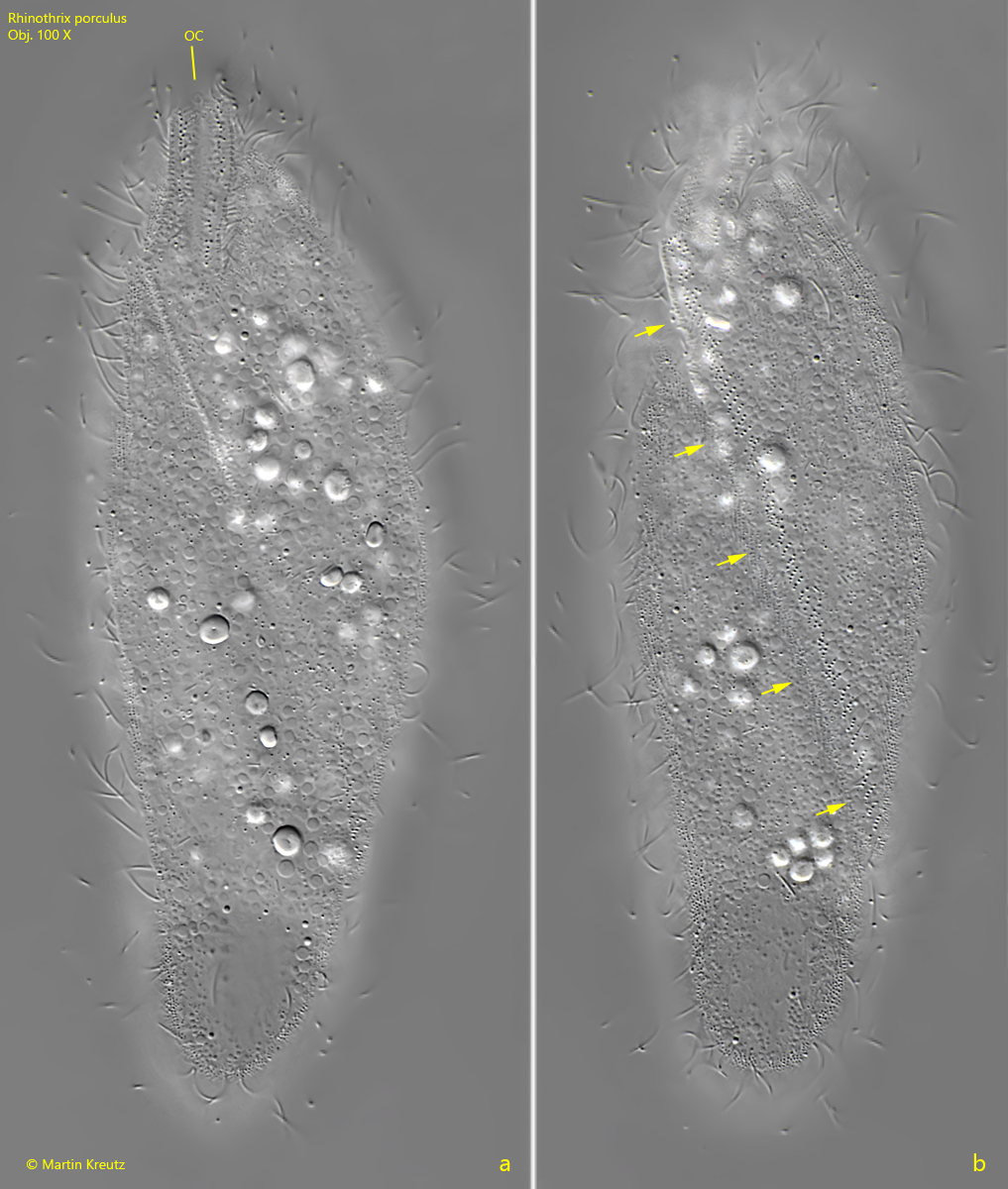
In slightly squashed specimens, the three-rowed dorsal brush is clearly visible to the right of the proboscis (s. fig. 5 a-b). On the ventral side, the oral bulge bends slightly to the left in the first quarter and then ends in a longitudinal bulge, which runs over the ventral side to the left and ends at the posterior end (s. figs. 2 c, 4 c and 9 a-b). It is covered with extrusomes of type 3.
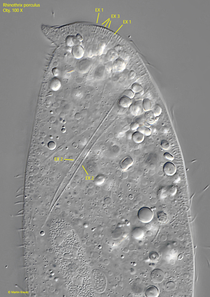
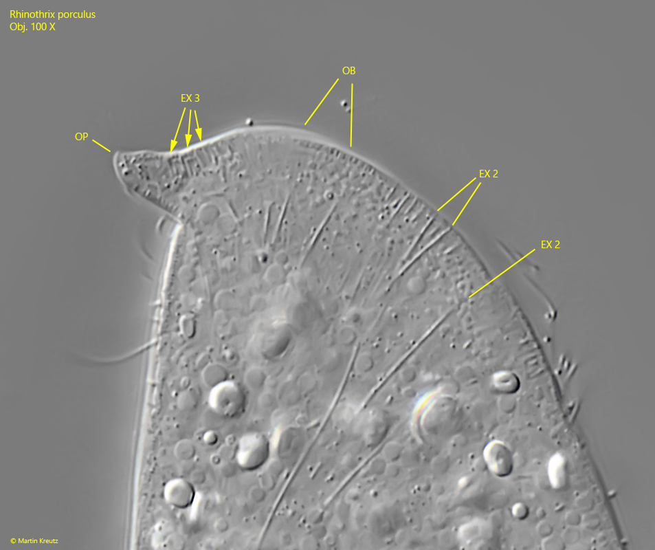
Rhinothrix porculus has 3 types of extrusomes (s. fig. 6). Between the ciliary rows of the body the curved extrusomes of type 1 are arranged, with a length of about 7 µm. In my specimens they were 6–7.4 µm long (s. figs. 6 and 8). The oral bulge and the oral palp is equipped with short, rod-shaped extrusomes of type 3 with a length of approx. 1.5 µm (s. figs. 6 and 7). In my specimens they were about 1.7 µm long (s. fig. 6). Finally, the extrusomes of type 2 are also attach to the oral bulge (s. fig. 7). They are slightly curved and about 50 µm long. However, they can also be found in the cytoplasm (s. fig. 6). In my specimens they were 38–45 µm long.
Some specimens of Rhinothrix porculus I found were over 160 µm long (s. fig. 2 a-d), which is larger than indicated by Foissner et al. Foissner carried out his measurements on 21 specimens impregnated with protargol. All specimens came from Simmelried samples that I sent to him in 2002. This can therefore only have been a small section of the population and changes in shape can occur during impregnation. I therefore believe that the larger specimens I found represent the common variability within the population.
Fig. 1 a-b:Rhinothrix porculus. L = 118 µm. A freely swimming (a) and slightly squashed specimen (b) from left found in March 2002. Note the oral bulge (OB) and the distinct oral palp (OP). Obj. 40 X.
Fig. 2 a-d:Rhinothrix porculus. L = 164 µm. A second, freely swimming specimen found in December 2022 from right (a), left (b) and from ventral (c, d). Obj. 40 X.
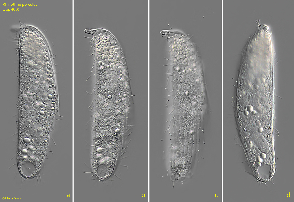
Fig. 3 a-d:Rhinothrix porculus. L = 138 µm. A third, freely swimming specimen, found in June 2023 from left (a-c) and from dorsal (d). Obj. 40 X.
Fig. 4 a-d:Rhinothrix porculus. L = 138 µm. The slightly squashed specimen as shown in fig. 3 a-d from left (a, b) and from ventral (c, d). Obj. 60 X.
Fig. 5 a-b:Rhinothrix porculus. Two focal planes of the three rowed dosal brush (DB). Obj. 60 X.
Fig. 6:Rhinothrix porculus. The three types of extrusomes (EX 1–3). The extrusomes type 1 are located between the ciliary rows and scattered over the body, while the extrusomes type 2 and type 3 are part of the oral bulge. In this specimen the extrusomes type 1 are 6 µm long, type 2 are 39-42 µm long and type 3 are 1.7 µm long. Obj. 100 X.
Fig. 7:Rhinothrix porculus. The oral palp (OP) in detail. Note the short extrusomes of type 3 (EX 3) in th oral palp what means it is a part of the oral bulge (OB). Some of the long extrusomes of Type 2 (Ex 2) are also inserted in the oral bulge. Obj. 100 X.
Fig. 8:Rhinothrix porculus. Focal plane on the curved extrusomes of the type 1 (EX 1). In this specimen they have a length of 6.9–7.4 µm. DB = dorsal brush, Ma = macronucleus, Mi = micronucleus. Obj. 100 X.
Fig. 9 a-b:Rhinothrix porculus. Two focal planes of the ventral bulge equipped with extrusomes type 3 as extension of the right side of the oral bulge. This longitudinal bulge running over the ventral side to the posterior end of the left side (arrows). OC = oral cleft. Obj. 100 X.
Fig. 10:Rhinothrix porculus. A squashed specimen with focal plane on the oblong macronucleus (Ma) and the attached hemi-spherical micronucleus (Mi). CV = contractile vacuole. Obj. 100 X.