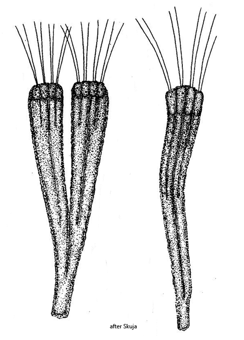
colonies dichotomous, loosly bushy, up to 2 mm long
branches of colony consist of 2–4 parallel fused tubes
each tube is covered with brownish or orange-brownish, hollow spherules
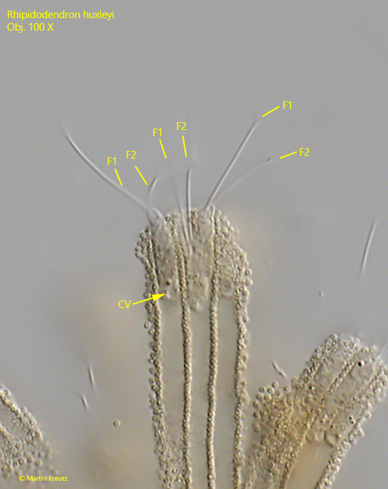
at distal end of each tube a 6–12 µm long, elongate oval flagellate
cells with 2 flagella of equal length, 2–3 times longer than cell
nucleus at anterior end
one contractile vacuole at posterior end
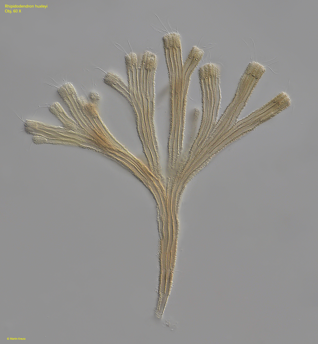
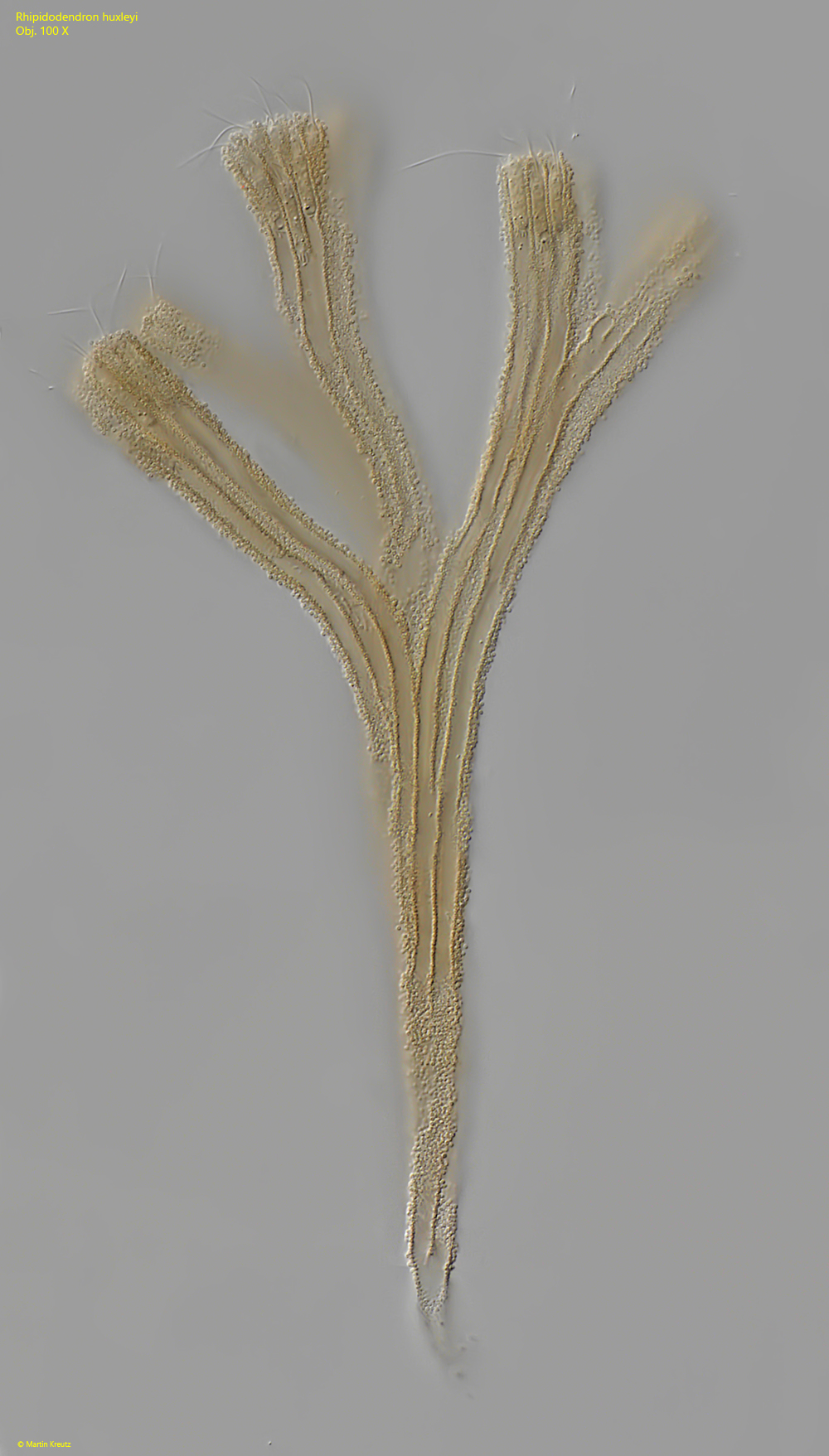
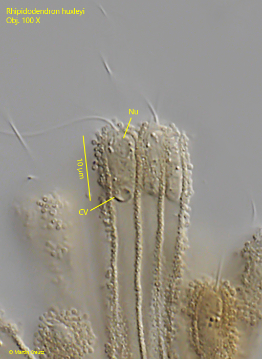
Rhipidodendron huxleyi
I find Rhipidodendron huxley very frequently in the Simmelried. In fresh samples it is difficult to examine the colonies because they are often covered with detritus and plant parts. However, Rhipidodendron huxley likes to settle on floating coverslips and can then be examined without disturbing foreign bodies.
The colonies of Rhipidodendron huxleyi have a very characteristic appearance due to their dichotomous branching and mostly brownish color. The colonies can also become very large with about 50 branches. Rhipidodendron huxleyi differs from the similar species Rhipidodendron splendidum in the structure of the branches. While in Rhipidodendron huxleyi these are made up of a maximum of 4 parallel tubes, in Rhipidodendron splendidum there can be considerably more tubes which also widen fan-shaped towards the distal end.
The cells at the end of the tubes have obviously been rarely studied because I have found very little information about them. The cells have two flagella of equal length and quickly withdraw into the tubes when slightly disturbed. The brownish globules covering the tubes make it difficult to examine the cells, especially as they denature and swell even under light coverslip pressure. According to my examinations, there is a spherical nucleus at the anterior end and one contractile vacuole at the posterior end.
The origin of the 0.6-0.7 µm spherules covering the tubes was investigated in more detail in the species Rhipidodendron splendidum (Hibbert, 1976). It was assumed that they were the remains of excreted food vacuoles. However, the spherules are hollow, except for one or sometimes two bacteria that are trapped inside. This can only be seen under an electron microscope. The spherules are formed in special vacuoles in which one bacterium at a time is surrounded by a brownish shell. The purpose of the enclosed bacteria is unknown.
Fig. 1:Rhipidodendron huxleyi. L = 260 µm (of colony). Total view of a medium sized colony. Note that a maximum of 4 flagellates sit next to each other at the distal end of the branches. Obj. 60 X.
Fig. 2:Rhipidodendron huxleyi. L = 210 µm (of colony). Total view of a smaller colony. Obj. 100 X.
Fig. 3:Rhipidodendron huxleyi. The distal end of a branch with 3 cells in detail. In the left cell the nucleus (Nu) is visible at the anterior end and the contractile vacuole (CV) at the posterior end. Obj. 100 X.
Fig. 4:Rhipidodendron huxleyi. The same cells as shown in fig. 3 with focal plane of the flagella. Each cell has two flagella (F1, F2) of equal length. CV = contractile vacuole. Obj. 100 X.