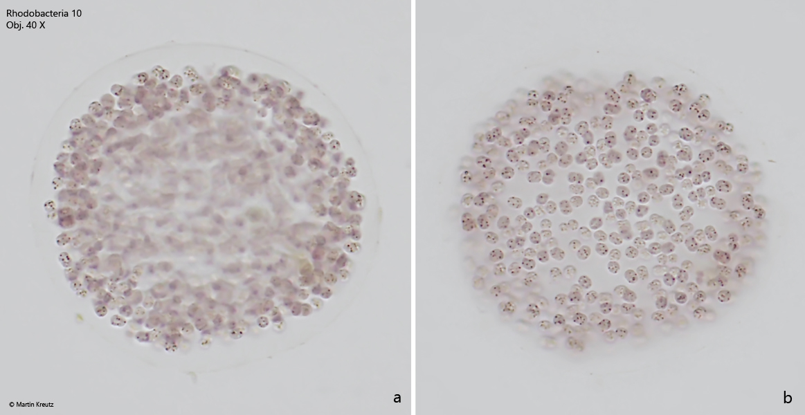
spherical shaped colonies, 50 – 100 µm in diameter
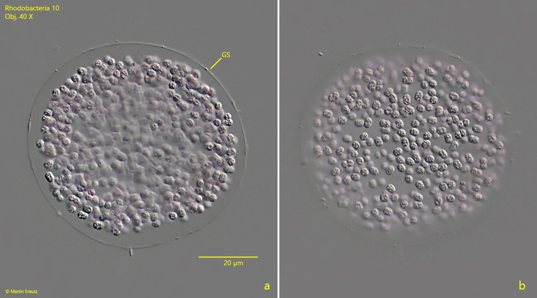
colonies are covered by a sharply defined gelatinuous sheat
cells in the colonies are separated from each other
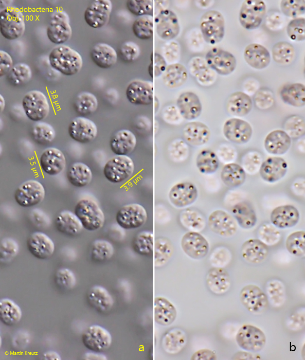
higly refractive spherules scattered in the cells
No drawings from previous authors available.
Fig. 1:Rhodobacteria 10. L = 3.4 – 3.9 µm. A slightly squashed colonie in brightfield illumination. All cells are separated from each other. Obj. 40 X.
Fig. 2:Rhodobacteria 10. L = 3.4 – 3.9 µm. The same colony shown in fig. 1 but in DIC. Not the sharply defined gelatinuous sheat (GS). Obj. 40 X.
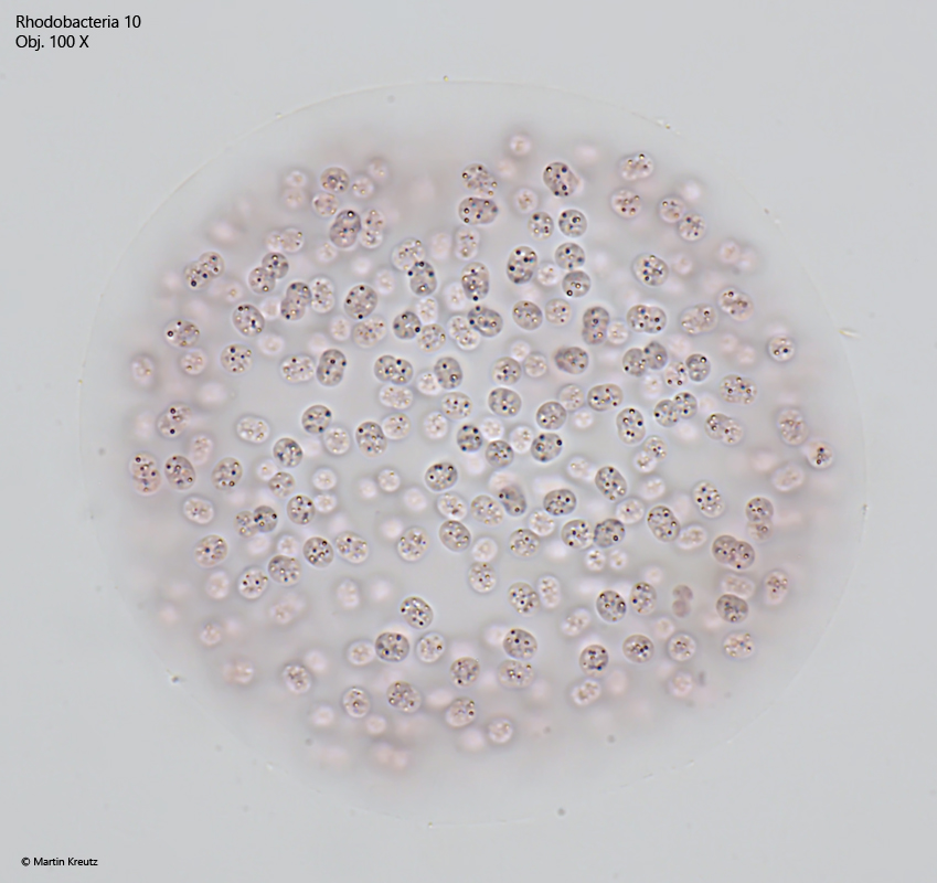
Fig. 3:Rhodobacteria 10. L = 3.4 – 3.9 µm. The cells in a squashed colony in brightfield illumination. The cells are slightly pink colored. Obj. 100 X.
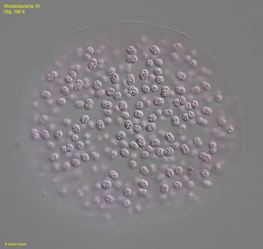
Fig. 4:Rhodobacteria 10. L = 3.4 – 3.9 µm. The same colony shown in fig. 3 but in DIC. Obj. 100 X.
Fig. 5 a-b:Rhodobacteria 10. L = 3.4 – 3.9 µm. The cells in a squashed colony in DIC (a) and brightfield illumination (b). Note the highly refracive spherules scattered in the cells. Obj. 100 X.