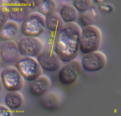
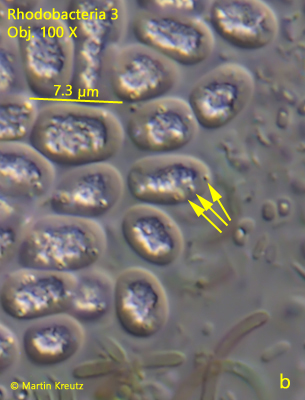
the cells are oval, larger cells oblong, sometimes irregularly
length 4 – 7.5 µm
center of the cells filled with irregularly shaped, highly refractive mass
slightly pink to intense pink, sometimes yellowish
colonies irregularly shaped without sharp outline
no visible gelatinuous sheat
colonies of about 50 – 250 µm in diameter
No drawings from previous authors available.
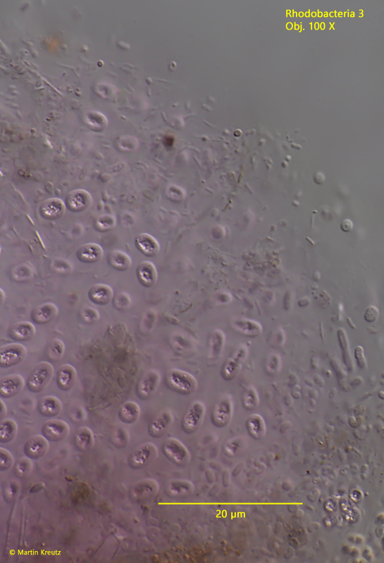
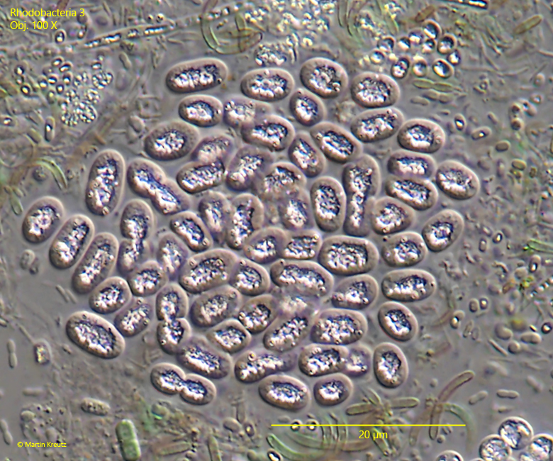
Fig. 1:Rhodobacteria 3. L = 4 – 7.5 µm. Part of a slightly squashed colony. All cells are separated from each other. Obj. 100 X.
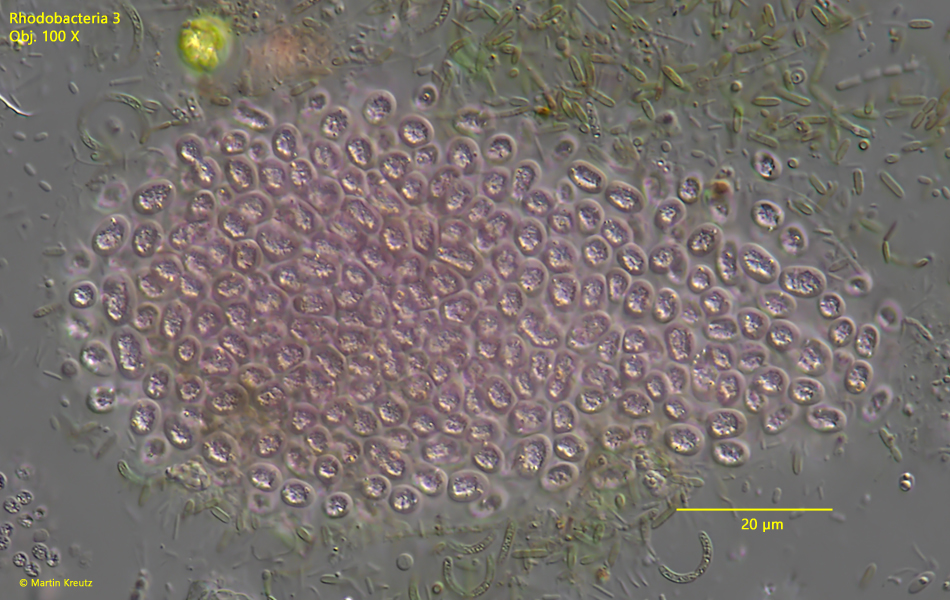
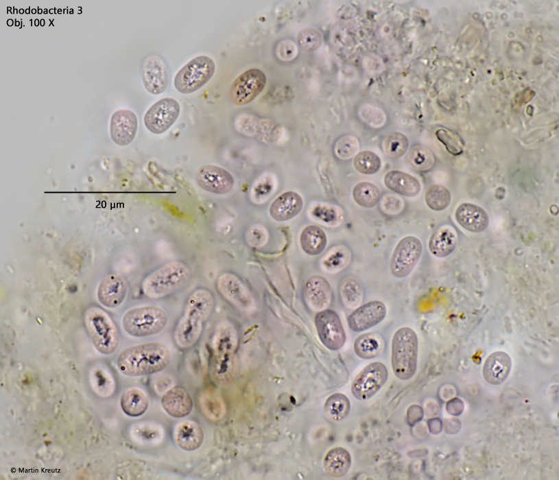
Fig. 2:Rhodobacteria 3. L = 4 – 7.5 µm. A squashed colony. The cells are embedded in a gelatinuous mass without sharp outline. Obj. 100 X.
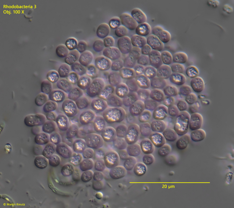
Fig. 3:Rhodobacteria 3. L = 4 – 7.5 µm. A squashed colony. Obj. 100 X.
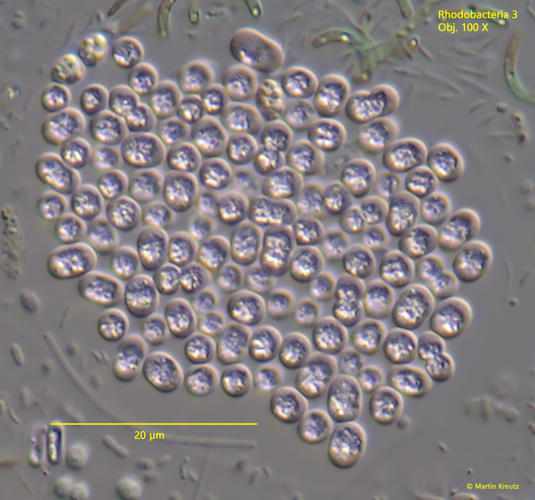
Fig. 4:Rhodobacteria 3. L = 4 – 7.5 µm. A squashed colony. Obj. 100 X.
Fig. 5:Rhodobacteria 3. L = 4 – 7.5 µm. A squashed colony with larger cells. Obj. 100 X..
Fig. 6:Rhodobacteria 3. L = 4 – 7.5 µm. A slightly squashed colony in brightfield illumination. Obj. 100 X.
Fig. 7 a-b:Rhodobacteria 3. L = 4 – 7.5 µm. The cells in detail. a) In some cells separate spherules are visible instead of the central highly refractive mass. b) In larger cells yellowish droplets are visible in the plasm (arrows) separated from the central highly refractive mass. Obj. 100 X.