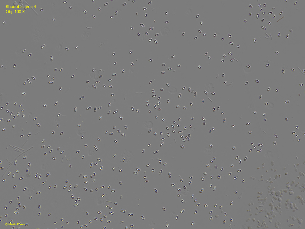
Fig. 1:Rhodobacteria 4. L = 2 – 3 µm. A slightly squashed colony in DIC. Obj. 100 X.
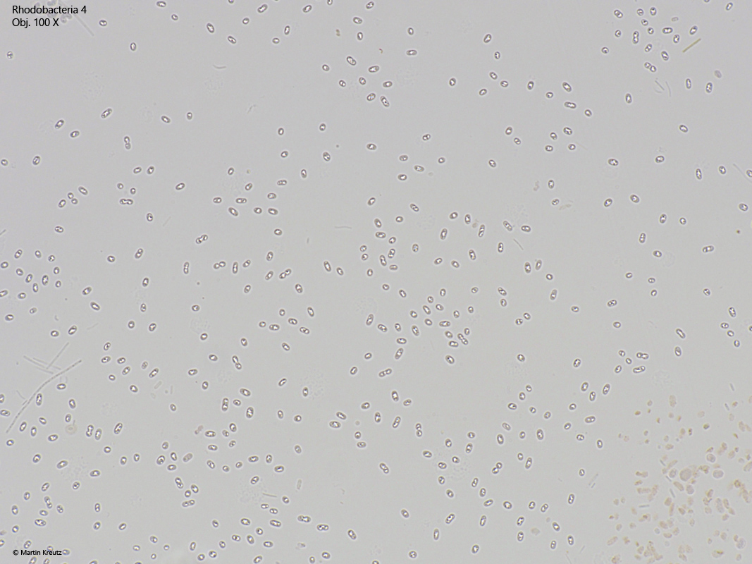
Fig. 2:Rhodobacteria 4. L = 2 – 3 µm. The same colony and the same image section as in fig. 1, but in brightfield illumination. Obj. 100 X.
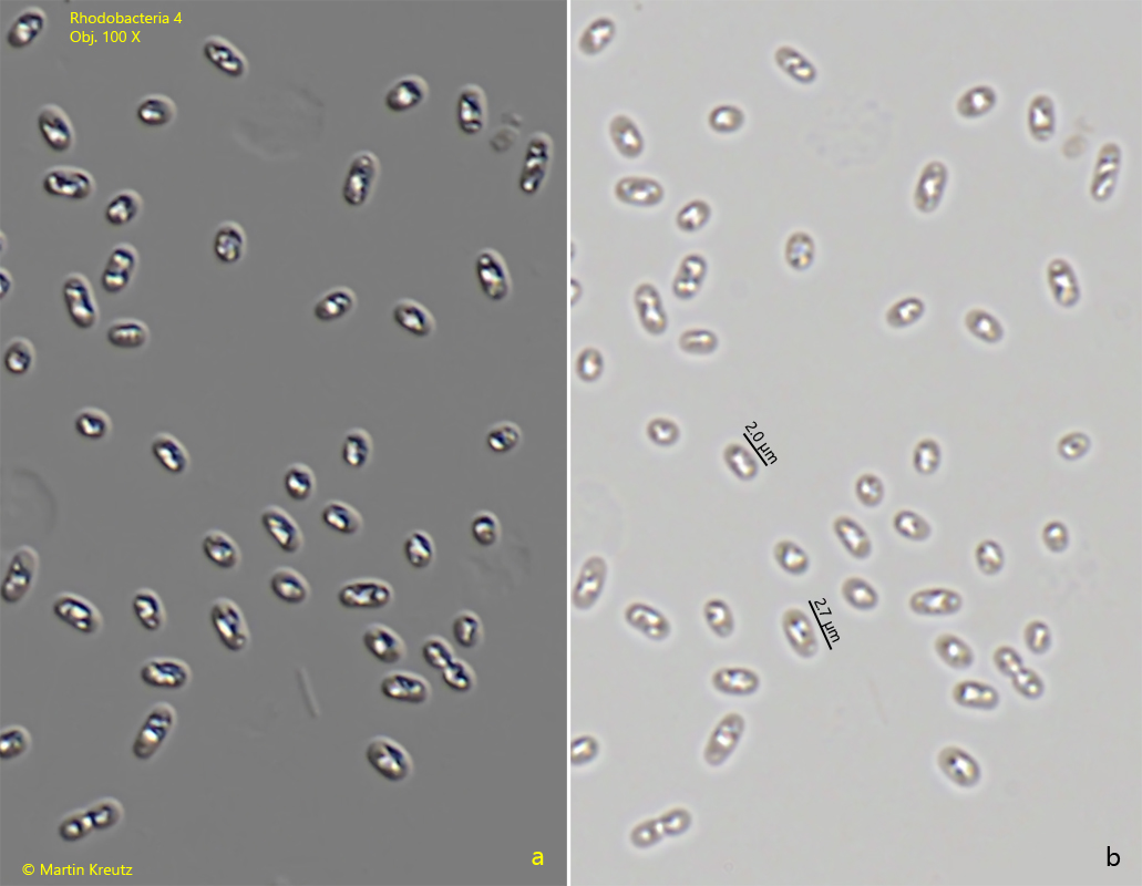
Fig. 3:Rhodobacteria 4. L = 2 – 3 µm. Comparison of the same enlarged sections from fig. 1 (DIC) and fig. 2 (brightfield). Obj. 100 X.
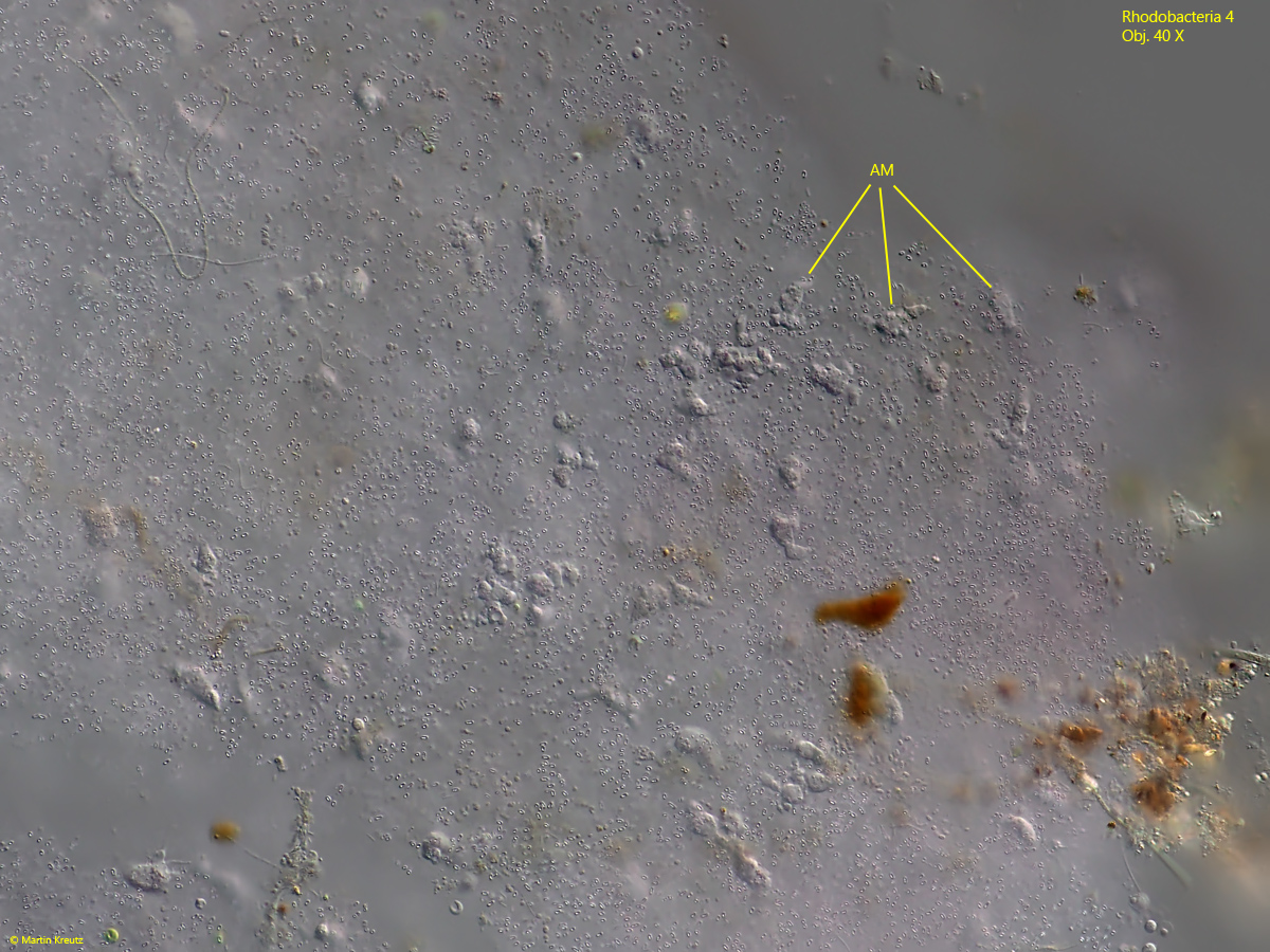
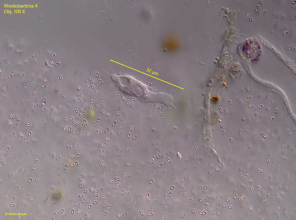
I found the colonies of rhodobacteria 4 often with an infestation of naked amoebae, which had settled on and in the colony and phagocytosed the rhodobacteria (s. Fig. 4). This was easily recognized by the pink food vacuoles in the amoebae. Obviously, these amoebae were specialized for this type of rhodobacteria, because I found them exclusively in the colonies of rhodobacteria 4. The amoebae are about 20 – 30 µm in size (s. Fig. 5). I was not able to classify these amoebae. However, I think it is possible that they represent an undescribed species, because amoebae specialized on Rhodobacteria have not been described so far to my knowledge.
Fig. 4:Rhodobacteria 4. A colony of rhodobacteria 4 infested with naked amoeba. AM = naked amoeba. Obj. 40 X.
Fig. 5:Rhodobacteria 4. One of the naked amoebae crawling on the colony in detail. Note the pink food vacuoles of the amoeba with remains of the refractive mass in the rhodobacteria. Obj. 100 X.