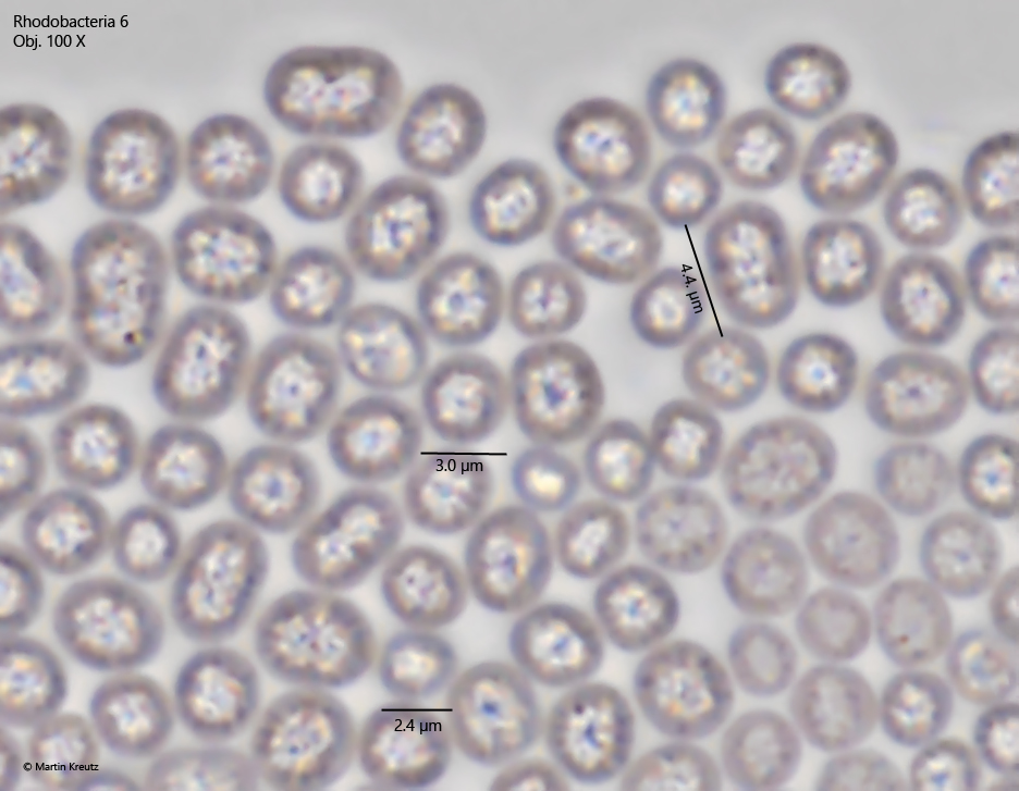
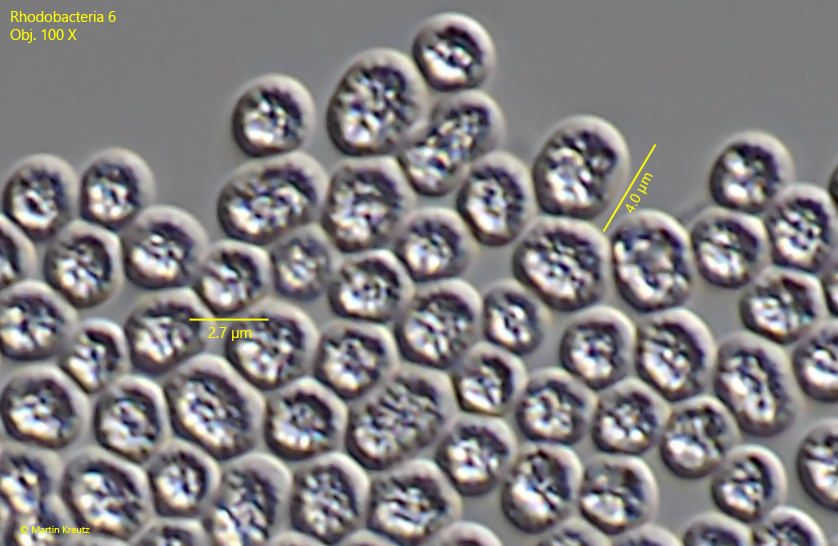
the cells are oblong, spherical and sometimes asymmetrically polygonal
length 2.4 – 4.5 µm
cells filled in the center with irregularly shaped, highly refractive mass
cells colored slightly pink or purple
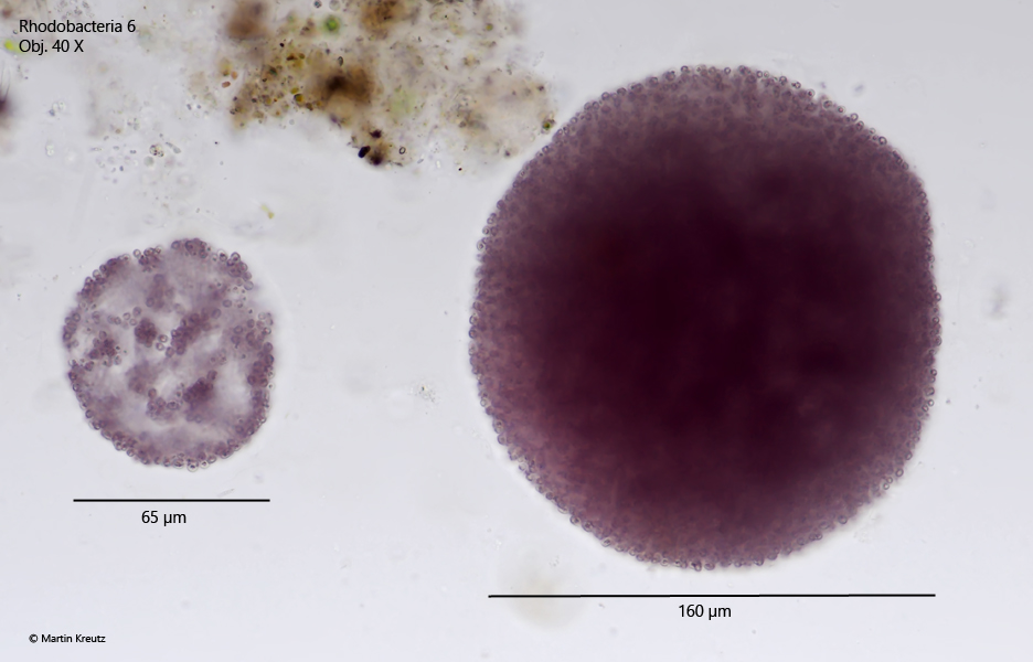
colonies round and spherical in shape, 30 – 250 µm in diameter
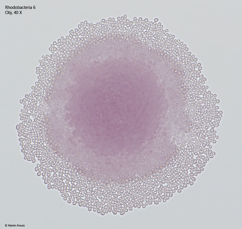
cells in the colonies are densely packed and touch each other
due to the high packing density, polygonal cells can be observed
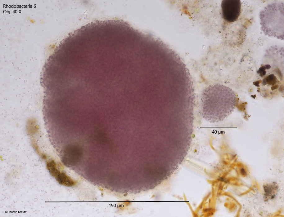
colonies are covered by a gelatinuous layer (hard to see)
colonies of about 30 – 250 µm in diameter
No drawings from previous authors available.
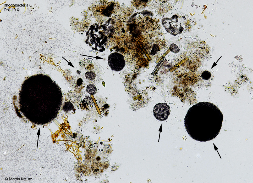
Fig. 1:Rhodobacteria 6. The spherical colonies appear almost black at low magnifications due to the high cell density (arrows). Obj. 10 X.
Fig. 2:Rhodobacteria 6. The spherical colonies appear distinctly pink or purple at higher magnification in brightfield illumination. Obj. 40 X.
Fig. 3:Rhodobacteria 6. Two further colonies. The larger colony is covered by a gelatinuous layer. Obj. 40 X.
Fig. 4:Rhodobacteria 6. The single cells in the colonies only become visible when they are strongly squashed. Obj. 40 X.
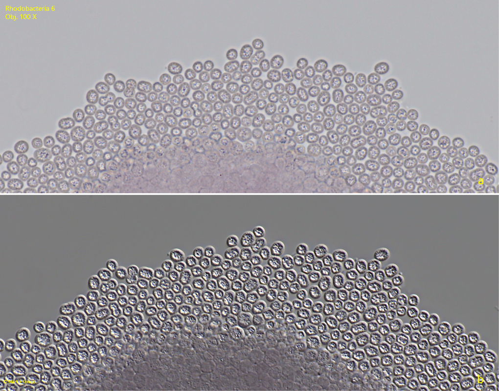
Fig. 5:Rhodobacteria 6. The cells in a squashed colony in brightfield illumination (a) and DIC (b). Obj. 100 X.
Fig. 6:Rhodobacteria 6. L = 2.4 – 4.5 µm. The cells in detail in brightfield illumination. The irregularly shaped mass is located in the center. The ring of cytoplasma around this mass appears darkly dottet in part or with dark shades. Obj. 100 X.
Fig. 7:Rhodobacteria 6. L = 2.4 – 4.5 µm. The cells in detail in DIC. Some of the cells are polygonally shaped. The central refractive mass fills the cells, except for the 0.5 µm thick fringe of cytoplasm. Obj. 100 X.