cells with highly refractive granules, mainly in the center
cells are colorless
colonies consist of loose aggregations of clusters of 2-16 cells
a gelatinuous sheat around the colonies, only visible due to adherent bacteria
colonies about 20 – 100 µm in diameter
No drawings from previous authors available.
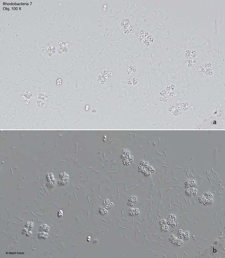
Fig. 1 a-b:Rhodobacteria 7. D = 1.8 – 2.2 µm. A colony of clustered cells in brightfield illumination (a) and DIC (b). Note the rod-shaped bacteria adhering to the gelatinuous sheat of the colony. Obj. 100 X.
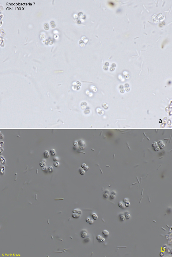
Fig. 2 a-b:Rhodobacteria 7. D = 1.8 – 2.2 µm. A second colony of clustered cells in brightfield illumination (a) and DIC (b). Obj. 100 X.
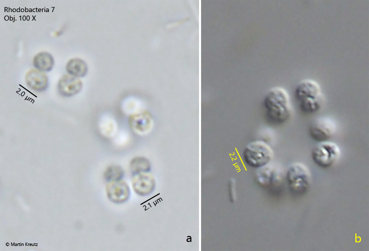
Fig. 3 a-b:Rhodobacteria 7. D = 1.8 – 2.2 µm. The cells of in detail in brightfield illumination (a) and DIC (b). Obj. 100 X.