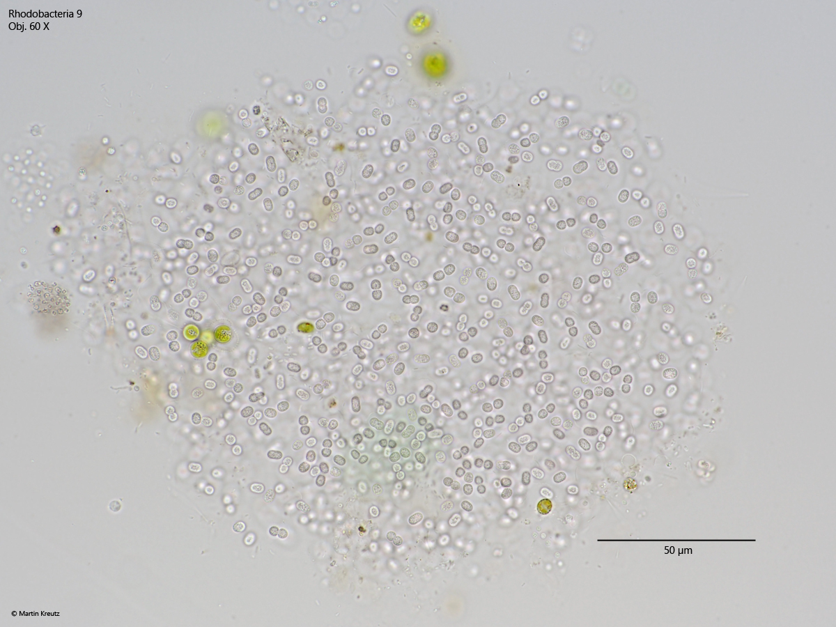
irregularly shaped colonies of about 50 – 250 µm in diameter
cells in the colonies are separated from each other
no visible gelatinous sheath
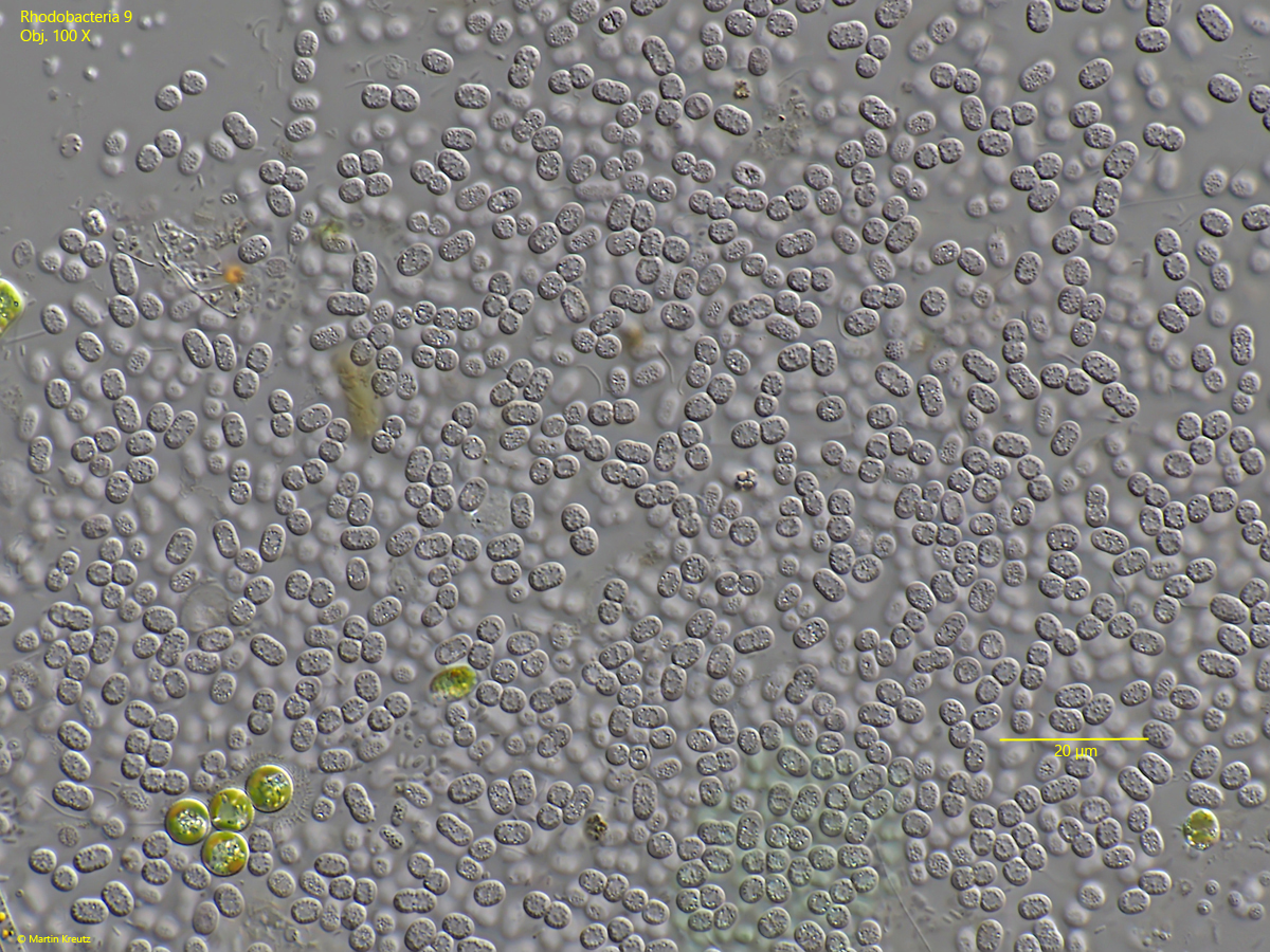
many division stages in the colonies visible
granules in the cells are arranged in a ring-shaped manner
No drawings from previous authors available.
Fig. 1:Rhodobacteria 9. L = 4.6 – 5.0 µm. A slightly squashed colony in brightfield illumination. All cells are separated from each other. Obj. 60 X.
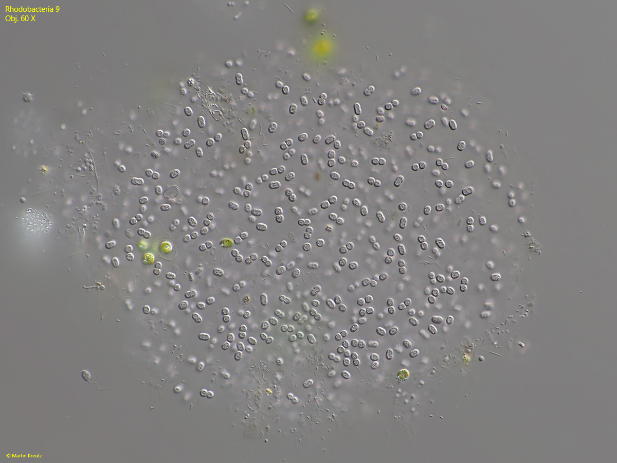
Fig. 2:Rhodobacteria 9. L = 4.6 – 5.0 µm. The same colony shown in fig. 1 but in DIC. Obj. 60 X.
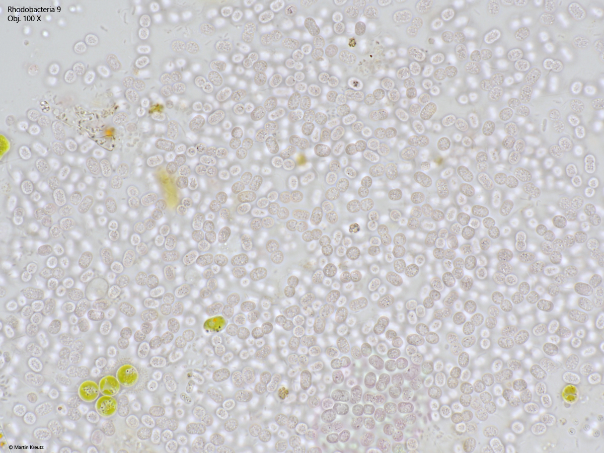
Fig. 3:Rhodobacteria 9. L = 4.6 – 5.0 µm. The cells in a squashed colony in brightfield illumination. The cells have a flesh-like color. Obj. 100 X.
Fig. 4:Rhodobacteria 9. L = 4.6 – 5.0 µm. The same field of view as shown in fig. 3 but in DIC. Note the high number of cells in the state of cell division (= “paired” cells). Obj. 100 X.
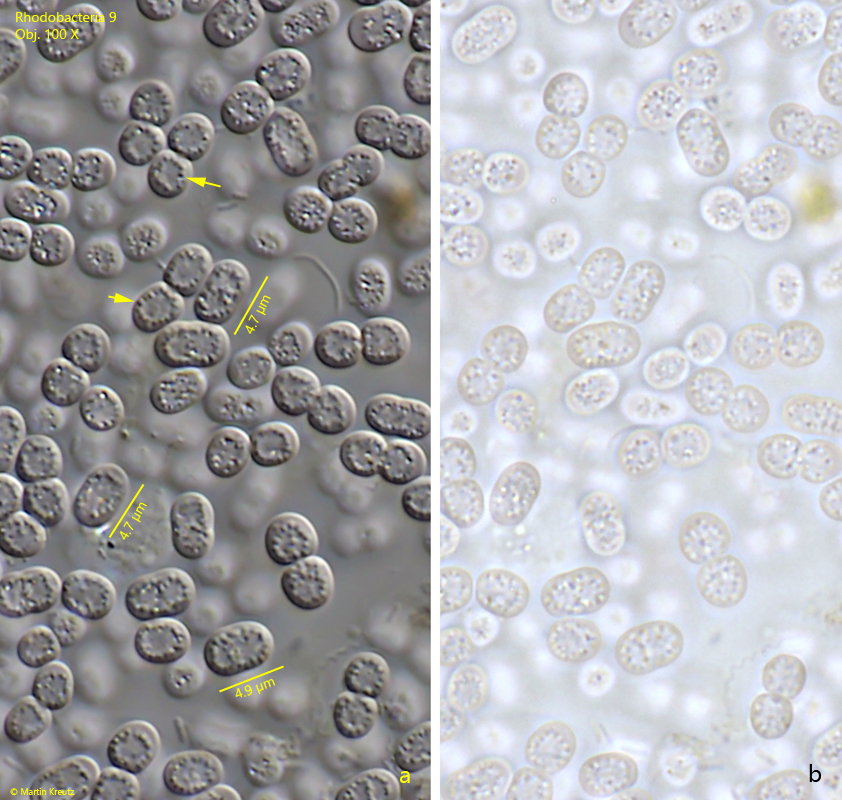
Fig. 5 a-b:Rhodobacteria 9. L = 4.6 – 5.0 µm. The cells in a squashed colony in DIC (a) and brightfield illumination (b). Note the granules arranged in a ring (arrows) while the center of the cells is almost free of granules. Obj. 100 X.