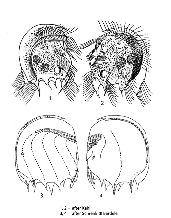
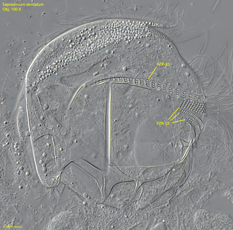
perizonal row on the right side reaches almost the dorsal keel, on left side short
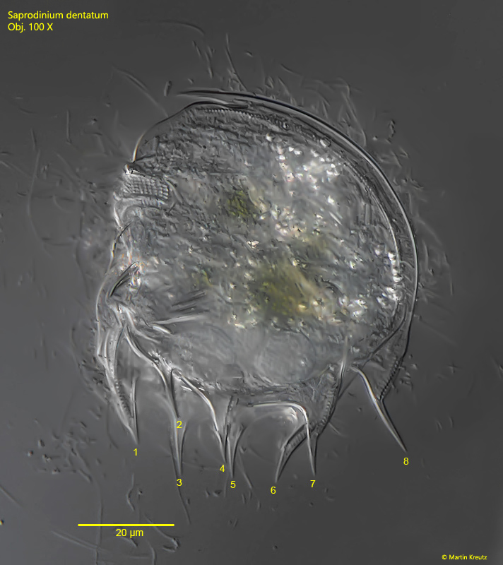
posterior indentation surrounded by 8 long spines
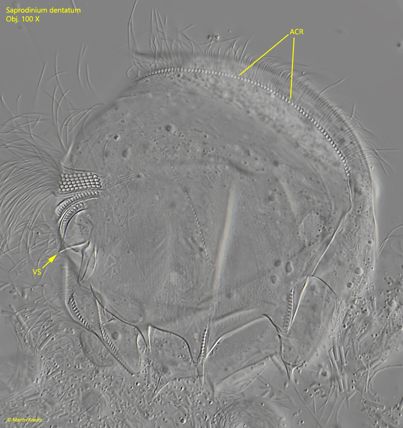
1 ventral spine below the perizonal row on the left side (hard to see)
3 macronuclei (rarely 2 macronuclei)
apical ciliary row, 2/3 of body length
short ciliary rows at distal end of spines, never reaching the cell equator
oral opening and adorale zone of membranelles in the middle of the ventral side
contractile vacuole below adorale zone of membranelles
Saprodinium dentatum
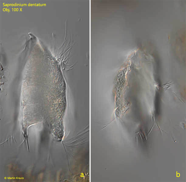
Saprodinium dentatum is by far one of the most common odontostomatid ciliates I find in my sites. The species is found in all waters with a mud layer with decaying leaves or between decaying plant masses. In contrast to other representatives of odontostomatid ciliates, Saprodinium dentatum is realtively large with 40–80 µm. In my populations the specimens were consistently longer than 70 µm and some specimens even reached 90 µm (s. fig. 3 a-b). The identification of this species is very simple. The body shape is mostly distinctly lenticular and there are 8 spines surrounding an indentation at the posterior end. There are 3 spines arranged on each side and two more on the anterior and posterior sides (s. fig. 4). The perizonal row is very long. It starts on the right side near the dorsal margin and covers the front side up to the left side, where it ends after a short distance (s. fig. 5). Characteristically, Saprodinium dentatum almost always has 3 macronuclei and only one micronucleus (s. figs. 2b and 3b). Like all odontostomatid ciliates, symbiotic bacteria are found also in Saprodinium dentatum (s. fig. 7). There are rod-shaped bacteria with straight ends (3.4–5.0 µm long) and rod-shaped bacteria with rounded ends (5.0–5.1 µm long).
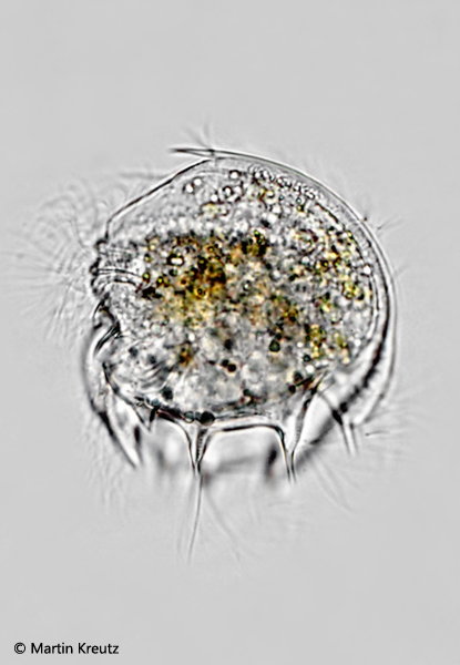
Fig. 1: Saprodinium dentatum. L = 74 µm. A freely swimming specimen from the left side in brightfield illumination. Obj. 40 X.
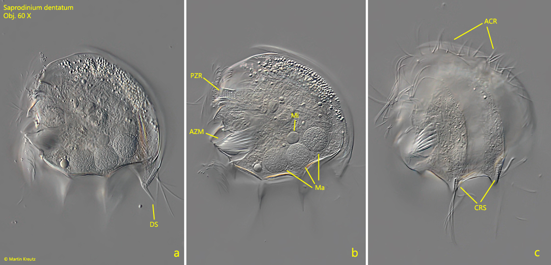
Fig. 2 a-c: Saprodinium dentatum. L = 71 µm. A freely swimming specimen from the left side. ACR = apical ciliary row, AZM = adoral zone of membranelles, CRS = ciliary row of the spines, Ma = macronuclei, Mi = micronucleus, PZR = perizonal row. Obj. 60 X.
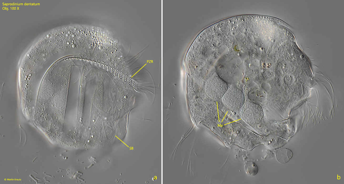
Fig. 3 a-b: Saprodinium dentatum. L = 90 µm. A slightly squashed specimen from the right side. Note the long perizonal row (PZR) on this side. Ma = macronuclei. Obj. 100 X.
Fig. 4: Saprodinium dentatum. A stack of 14 images from the left side to visualize the 8 posterior spines (1–8). The median spines nos. 2,3–4,5–6,7 are arranged in pairs, while ventrally (1) and dorsally (8) only 1 spine arises each. Obj. 100 X.
Fig. 5: Saprodinium dentatum. A stack of 2 images from the right side to visualize the perizonal row, running from the right side (PZR-RS) over the ventral side to the left side (PZR-LS). Note that the perizonal row on the right side is much longer. Obj. 100 X.
Fig. 6: Saprodinium dentatum. Strongly squashed specimen from the left side to visualize the small ventral spine (VS) below the perizonal row. This ventral spine is hard to see in a freely swimming specimen. ACR = apical ciliary row. Obj. 100 X.
Fig. 7: Saprodinium dentatum. The symbiotic bacteria released from a strongly squashed specimen. There are two types of bacteria. The rod-shaped bacteria with straight ends (SB1) are 3.4–5.0 µm long (during cell division apparently longer) while the second type of bacteria (SB2) has rounded ends and is 5–5.1 µm long. Obj. 100 X.
Fig. 8: Saprodinium dentatum. A freely swimmung specimen from dorsal (a) and posterior (2). Obj. 100 X.