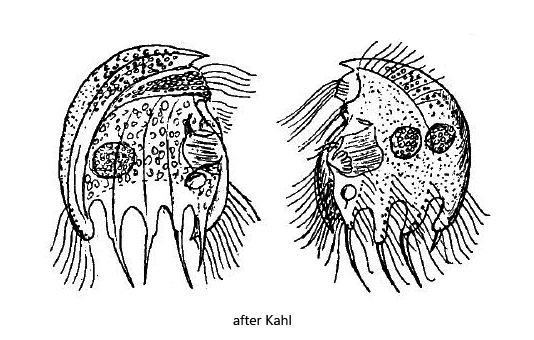
tooth of the right side without ciliary rows apart from the dorsal tooth
left side with 3 long spines and one short spine on the dorsal tooth
all tooth of the right side with ciliary rows
one or two macronuclei
oral opening and adorale zone of membranelles the middle of the ventral side
contractile vacuole below adorale zone of membranelles
Saprodinium integrum
I have found Saprodinium integrum several times in Simmelried since 1993, but always only single specimens. I have no evidence from my other sites. I found the species in both summer and winter months.
The identification of Saprodinium integrum is not easy, because determining the exact arrangement of the spines at the posterior is not simple, since one can look at a specimen under the cover glass only from one side. A good indication that of Saprodinium integrum are the spines of the left side bent backwards (s. fig. 2 a-d), explicitly mentioned by Kahl. It is helpful to squash a specimen strongly to have all spines in focus at the same time (s. fig. 3).
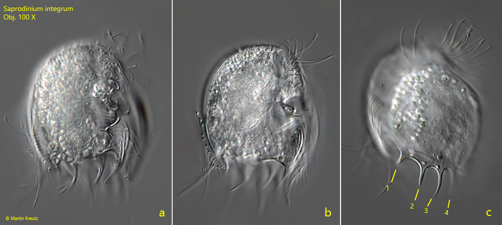
Fig. 1 a-c: Saprodinium integrum. L = 46 µm. A freely swimming specimen from the right side. The 4 long spines of the right side are visible in fig. c (1-4). The blurry spines in figs. a and b are the bent spines of the left side. Obj. 100 X.
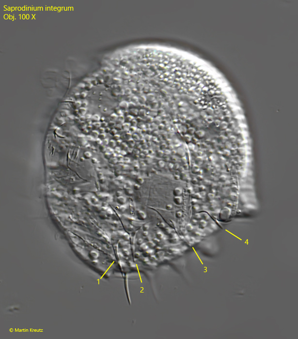
Fig. 2 a-d: Saprodinium integrum. L = 44 µm. A freely swimming specimen from the left side. On the left side 3 long spines (1–3) are visible and a short spine at the dorsal tooth (4). Note that all spines of the left side are bent backwards. AZM = adorale zone of membranelles, CR = ciliary rows of the tooth, Ma = macronuclei, PCR = perizonal ciliary row. Obj. 100 X
Fig. 3: Saprodinium integrum. A squashed specimen from the left side. The long spines are visible (1-3) and the short spine (4) of the dorsal tooth. Obj. 100 X