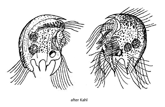
right between ventral tooth and dorsal spine two spines
left two spines and the dorsal spine
prominent frontal tooth
two spherical macronuclei, each with adjacent micronucleus
contractile vacuole in posterior third below AZM
Saprodinium mimeticum
So far I found Saprodinium mimeticum exclusively in the Simmelried. The species occurs there only very rarely. The photographs shown below are from 3 specimens I found between March and June 2008. After that I have not found the species again. Members of the genus Saprodinium can be recognized by the dorsal spine, which in many species is also directed dorsally (s. fig. 2b). A dorsal spine is present on the right as well as on the left side. The species Saprodinium mimeticum can be recognized by the ventral tooth is large and curved and also does not bear a spine (s. figs. 1a and 2a). Moreover, this large ventral tooth which is found only on the right side. On the left side, it is absent or strongly reduced (s. fig. 3b). There are also two more spines on each side at the posterior end (SP1 and SP2, s. fig. 3b). On the left side, Saprodinium mimeticum thus has these 2 spines at the posterior end, as well as the dorsal spine. On the right side one finds the prominent ventral tooth, the two middle spines and the dorsal spine.
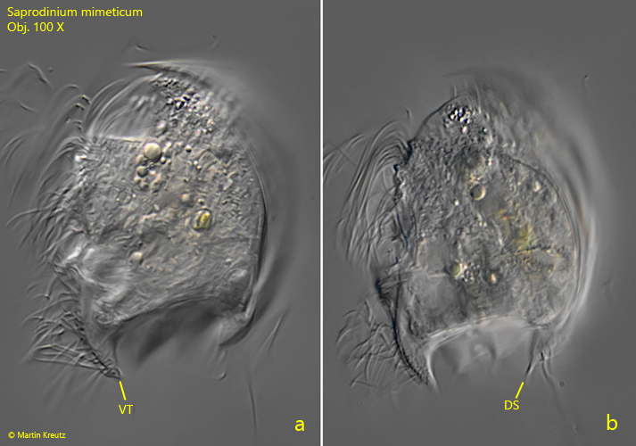
Fig. 1 a-b:Saprodinium mimeticum. L = 41 µm. Lateral view of the right side of a freely swimming specimen. Note the prominent and curved ventral tooth (VT) lacking a spine. DS = dorsal spine. Obj. 100 X.
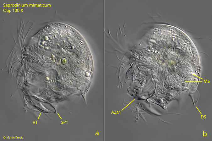
Fig. 2 a-b:Saprodinium mimeticum. L = 42 µm. Lateral view of the right side of a second freely swimming specimen. AZM = adoral zone of membranelles, DS = dorsal spine, Ma = macronuclei, SP1 = spine 1 on right side, VT = ventral tooth. Obj. 100 X.
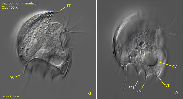
Fig. 3 a-b:Saprodinium mimeticum. L = 37 µm. Lateral view of the left side of a third freely swimming specimen. Note the strongly reduced ventral tooth (RVT) of the left side. CV = contractile vacuole; DS = dorsal spine; FT = frontal tooth; SP1, SP2 = spines of the left side. Obj. 100 X.