body ovoid shape, slightly tapered to posterior end
dorso-ventral slightly flattened
mouth opening apical, with a ventral mouth slit
circular oral bulge, mouth slit straight
row of 5–7 extrusome bundles from anterior to posterior on ventral side
extrusomes 50–58 µm long, flexible fibers
on average 102 ciliary rows
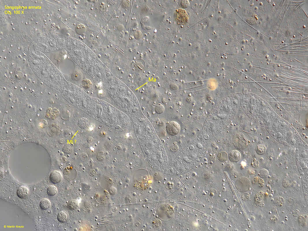
macronucleus ribbon-shaped
dorsally a field of papillae (5 rows)
dorsal brush 20 rows (hard to see)
contractile vacuole terminal
thickened pellicle with a fringe of 1–2 µm long extrusomes
Songophrya armata
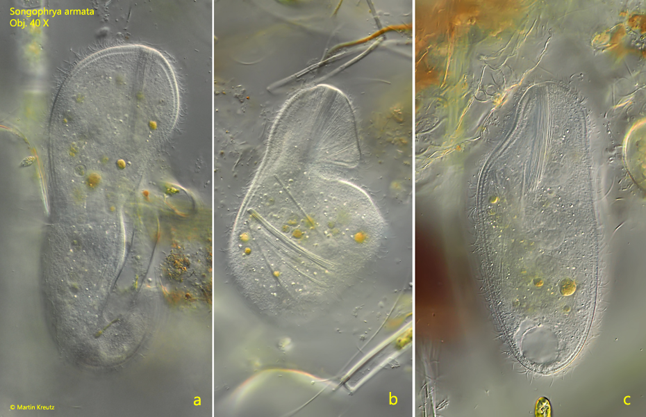
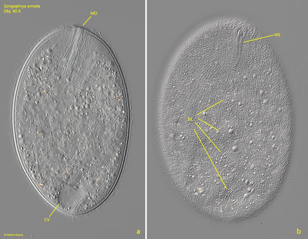
In April 2020 I noticed prostomatid ciliates in an old sample of decomposing plant material from Simmelried. At low magnifications I considered these ciliates to be Holophrya. But then I noticed that they were very metabolic and burrowed among the plant material (s. fig. 1 a-c) and that they were dorso-ventrally flattened (s. fig. 2 a-b). These characteristics did not match Holophrya. I isolated the 180–220 µm long specimens and examined them more closely at higher magnifications. I immediately noticed a ventral row of bundles of extrusomes extending from the mouth opening to the posterior end (s. figs. 4 and 5). There were between 6–7 of these bundles of extrusomes. The mouth opening was slightly displaced ventrally and surrounded by a circular oral bulge (s. fig. 8). Ventrally, there was also a straight, slit-shaped mouth opening, in the extension of which the bundles of extrusomes were arranged (s. fig. 5). On the dorsal side, immediately behind the mouth opening, there is a 5-row papillae field (s. figs. 6, 7 and 8). I could not clearly identify a dorsal brush. The macronucleus is ribbon-shaped (s. figs. 11 and 12). Probably only one spherical micronucleus is present, but I could not identify it with certainty (s. figs. 10 and 12). The contractile vacuole is located terminally (s. fig. 3a). The extrusomes are 50–58 µm long flexibles fibers (s. figs. 8 and 9). There is a distinctly thickened pellicle visible with a fringe of 1–2 µm long extrusomes (s. fig. 9). I identified the ciliate as Songophrya armata based on these characteristics. Later I realized that I had already photographed this species in August 2008 (also in a sample from Simmelried), but had failed to recognize it then. After the identification in April 2020 I could find Songophrya several times, always among rotting plants from the Simmlried. I could also notice that the species was especially abundant in old samples that were several weeks old. So far I have detected Songophrya armata exclusively in the Simmelried.
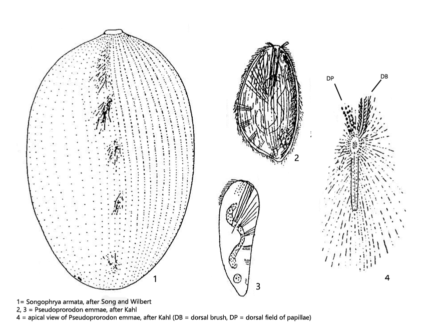
The history of the taxonomic classification of Songophrya armata is quite complex. When I attempt to assign the ciliate to a known species I came across Pseudoprorodon emmae (Berg, 1896) described and drawn by Kahl (s. drawings 2 and 3 above). Kahl also recognized and drew the strange dorsal field of papillae (s. drawing 4 above) of this ciliate and the bundles of extrusomes of the ventral side. However, the species I found has 5 rows of dorsal papillae (s. figs. 6, 7 and 8). Therefore I searched for a redescription of Pseudoprorodon emmae and found the description of the Myriokaryonidae by Foissner, 2003 (Foissner, 2003, s. Literature). In this article Foissner describe Holophrya (Pseudoprorodon) emmae and renamed it to Berghophrya emmae. In this article Foissner also shows a drawing of the ventral side with the bundles of extrusomes of Bergophrya emmae, which Kahl does not show. The bundles of extrusomes in this species are distributed over a larger field on the ventral side and they are not arranged in a row as in my ciliate. So my ciliate could not be Berghophrya emmae (= Holophrya emmae = Pseudoprorodon emmae) because of this feature and the differently constructed papilla field. However, a further ciliate is also described in the same article. This species was originally described by Song and Wilbert as Holophrya emmae. Based on the characteristics described by these authors, Foissner concluded that it is not Holophrya emmae, but a new species. This new species, identified solely on the basis of the description by Song and Wilbert, was named Songophrya armata by Foissner (Foissner, 2003). Foissner never found Songophrya armata himself. Due to this history the species Songophrya armata is not currently considered valid. In his article on the Myriokaryonidae, Foissner shows a drawing by Song and Wilber of the ventral side of Songophrya armata (s. drawing 1 above). The extrusomal bundles of this species are arranged in a straight longitudinal line, as I have seen in my species. In addition, I could see the circular mouth bulge around the mouth opening (s. figs. 6 and 8), which is also a characteristic of the genus Songophrya. Therefore I can confirm these characteristics on the basis of life observations, which were described by Song and Wilbert and which caused Foissner to establish a new species. Since there is also no meaningful alternative, I think the assignment Songophrya armata is correct.
Fig. 1 a-c:Songophrya armata. L = 220 µm. A specimen burrows among rotting plants under metabolic deformation. Obj. 40 X.
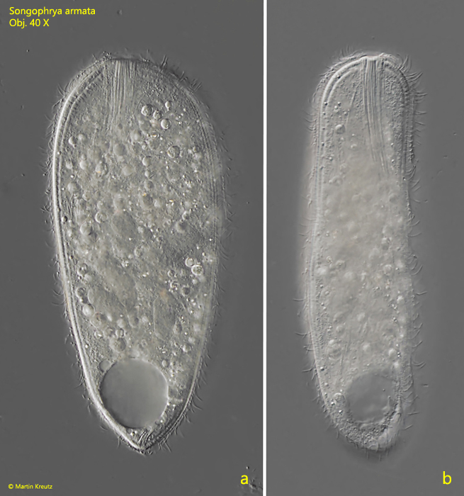
Fig. 2 a-b:Songophrya armata. L = 182 µm. A freely swimming specimen from ventral (a) and lateral from right (b). Obj. 40 X.
Fig. 3 a-b:Songophrya armata. L = 182 µm. Two focal planes of a slighty squashed specimen. On the ventral side (b) the row of extrusome bundles (BE) is visible. CV = contractile vacuole. Obj. 40 X.
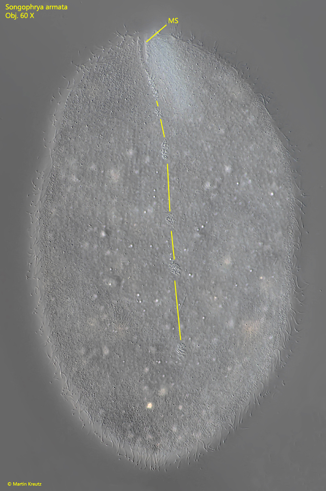
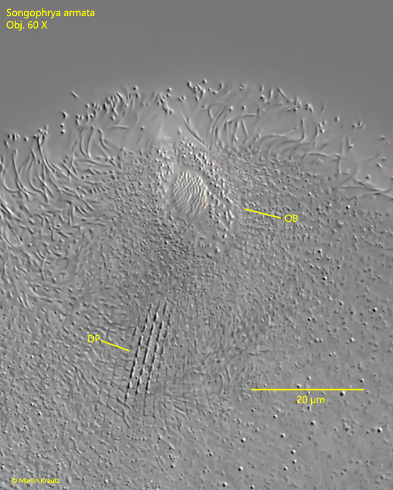
Fig. 4:Songophrya armata. L = 200 µm. A more detailed ventral view of the extrusome bundles running in a straight line from the apical mouth slit (MS) to the anterior end. Obj. 60 X.
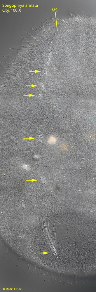
Fig. 5:Songophrya armata. The ventral row of bundles of extrusomes (arrows) in a second specimen. MS = mouth slit. Obj. 100 X.
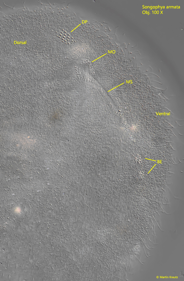
Fig. 6:Songophrya armata. This semi-apical view shows the arrangement of the dorsal field of papillae (DP) posterior to the mouth opening (MO) and the mouth slit running ventrally and the transition to the row of extrusomal bundles (BE) adjoining it. Obj. 100 X.
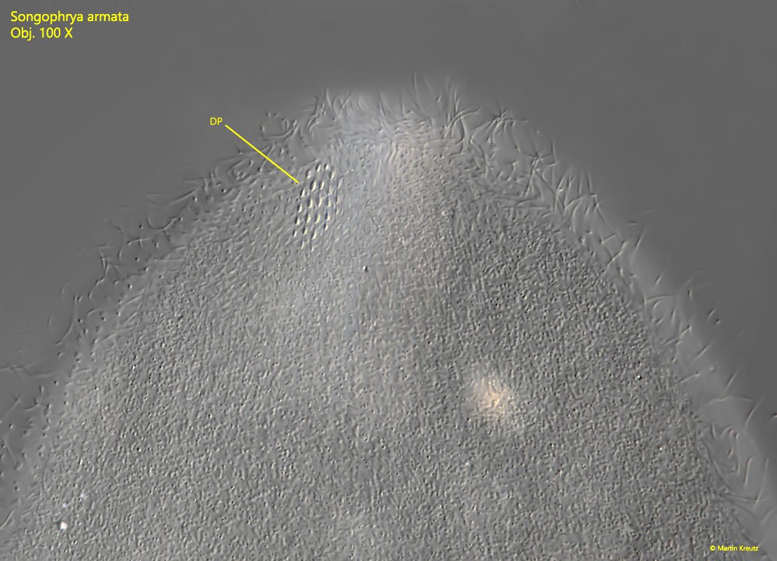
Fig. 7:Songophrya armata. The dorsal field of papillae (DP) in detail. It consists of 5 rows of 3–6 papillae each. Obj. 100 X.
Fig. 8:Songophrya armata. A dorsal view on the circular oral bulge (OB) and the dorsal field of papillae (DP). Obj. 100 X.
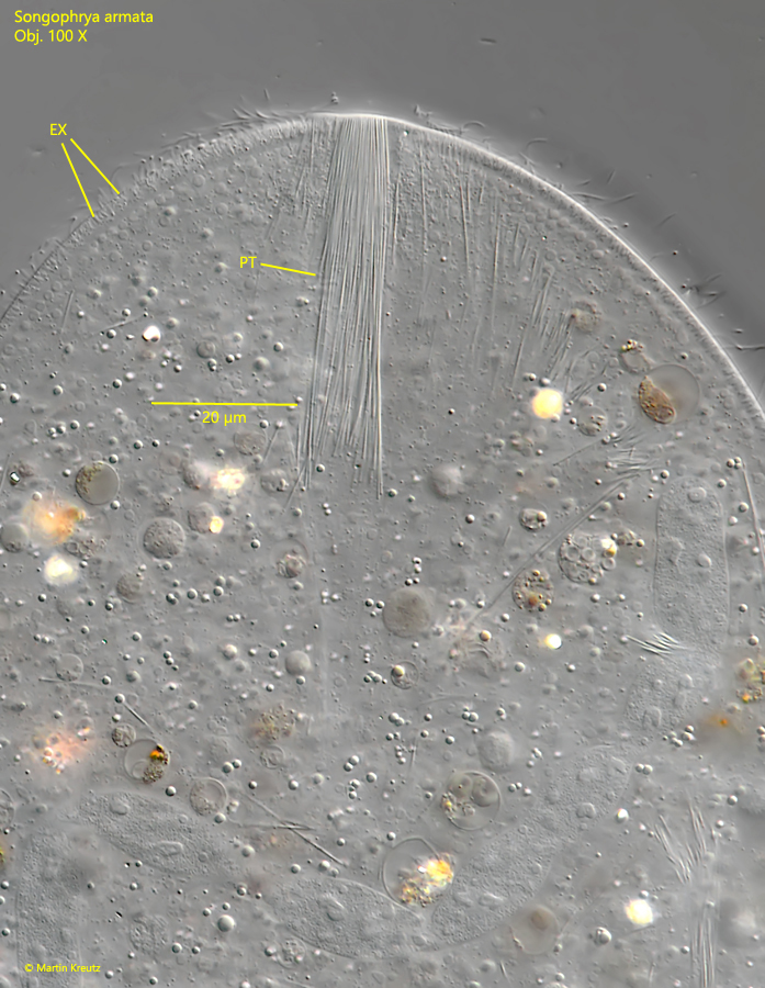
Fig. 9:Songophrya armata. The pharyngeal trichites (PT) in a squashed specimen. There is a fringe of 1-2 µm long extrusomes (EX) in the thickened pellicle. Obj. 100 X.
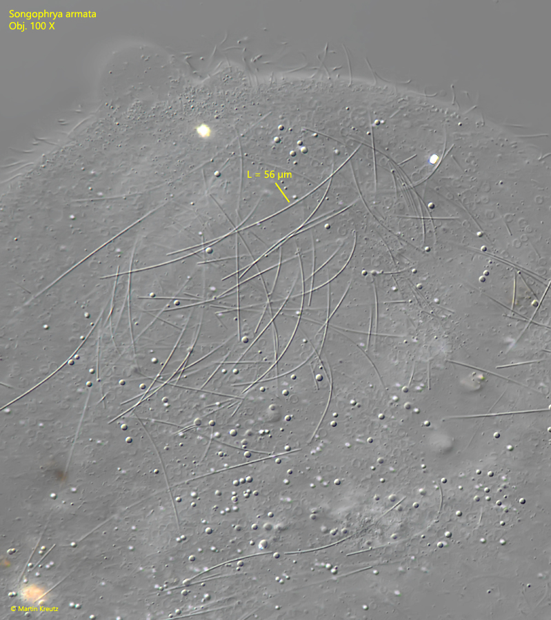
Fig. 10:Songophrya armata. The fiber-shaped, flexible extrusomes with a length of 50 – 58 µm in a strongly squashed specimen. Obj. 100 X.
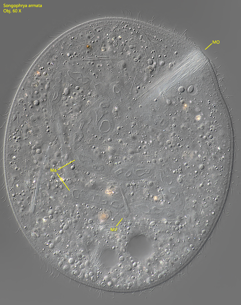
Fig. 11:Songophrya armata. A total view of a squashed specimen with the ribbon-shaped macronucleus (Ma). Mi? = probably the spherical micronucleus. Obj. 100 X.
Fig. 12:Songophrya armata. The ribbon-shaped macronucleus (Ma) in a second specimen. Mi? = probably the spherical micronucleus. Obj. 100 X.