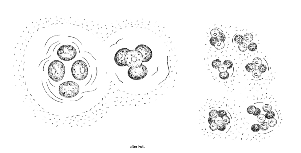
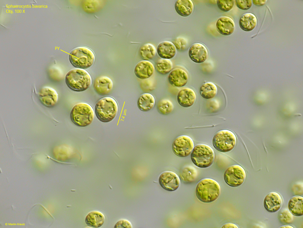
young cells almost spherical in tightly arranged groups of 2–4–8 cells
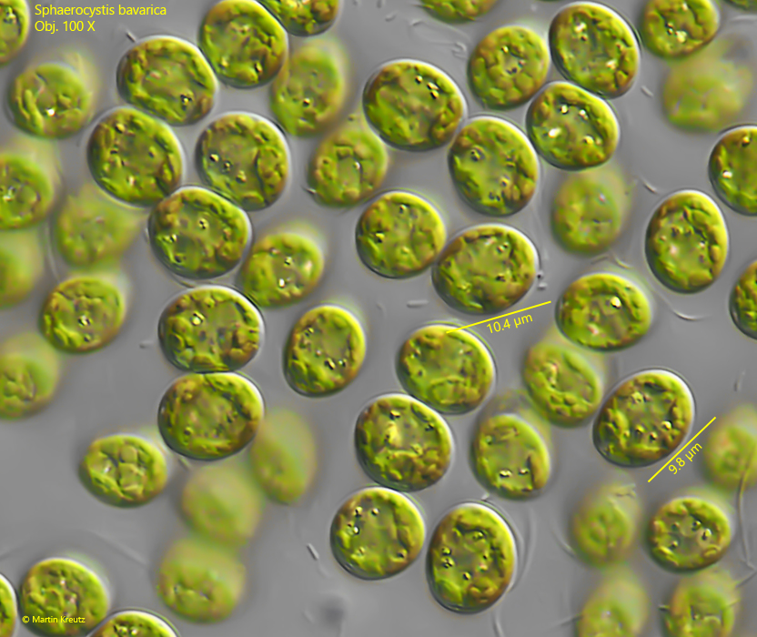
older cells broadly ovoid or ellipsoidal (6–15 µm long) in widely spaced groups
cell wall thin and smooth
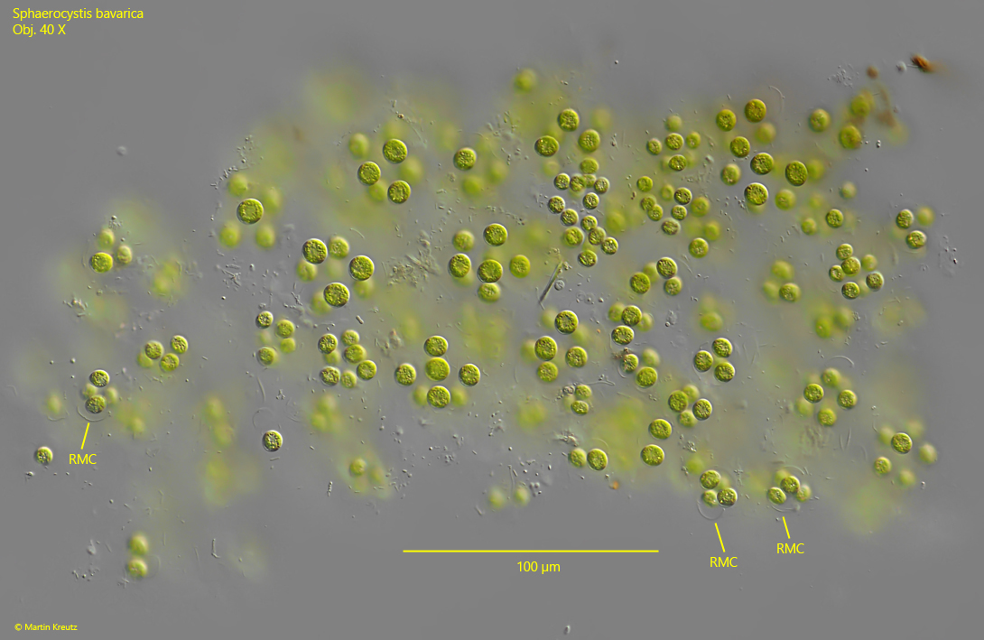
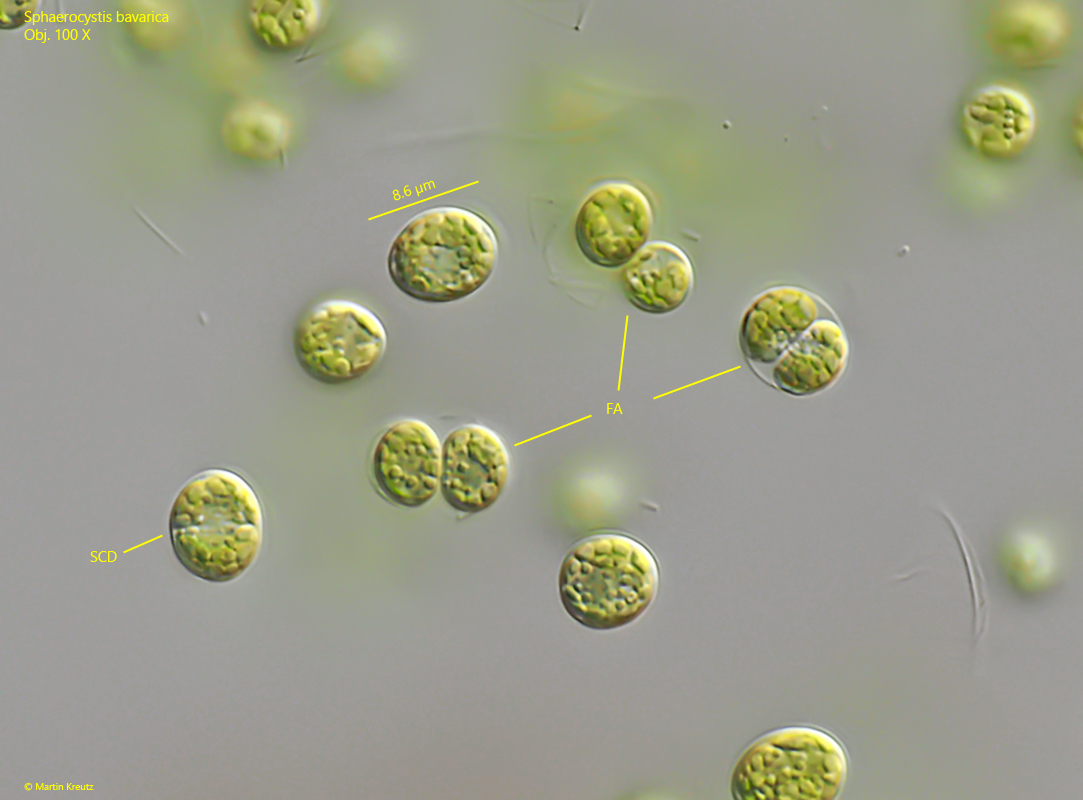
during autospore formation, the cell wall disintegrates into colonial mucilage
single, parietal chloroplast
one pyrenoid
asexual reproduction by autospores or zoospores with 2 apical flagella
benthic lifestyle
Sphaerocystis bavarica
Sphaerocystis bavarica is one of the most common chlorococcal algae in Simmelried. In particular in pond 6 it occurs in masses since this pond has started to become increasingly silted up.
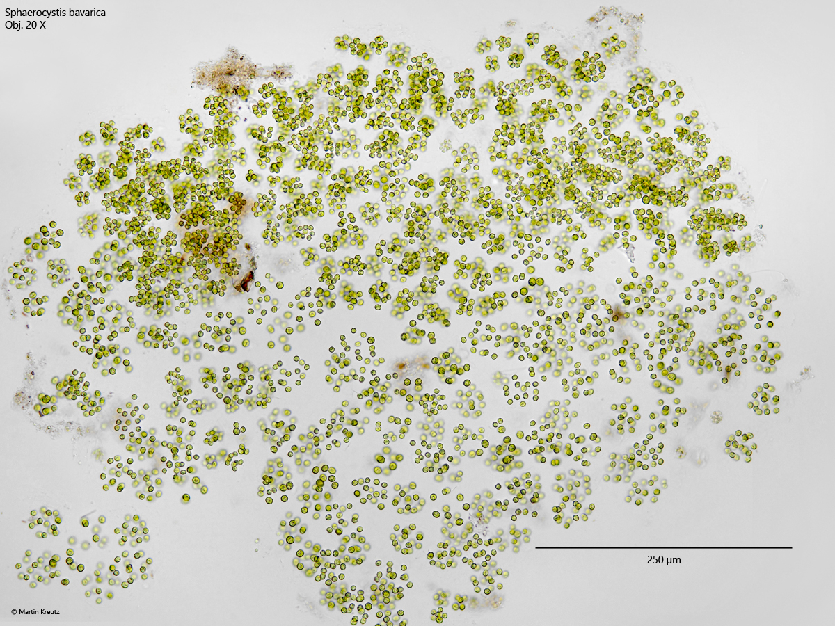
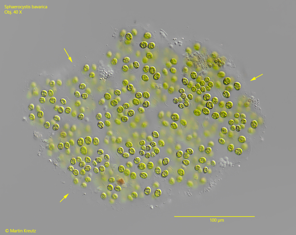
Sphaerocystis bavarica forms very large colonies that can contain more than 500 cells and are up to one millimeter in size. The colonies of Sphaerocystis bavarica are characterized by the fact that they do not have a sharply defined envelope and that the groups of cells within the colony consist of young, spherical cells as well as older, broadly ovoid cells. With increasing age, the cells move away from each other and the cell wall of the autospores dissolves. This gives the colony a heterogeneous character.
Sphaerocystis bavarica can easily be confused with Sphaerocystis schroeteri and Pseudosphaerocystis lacustris. However, Sphaerocystis schroeteri is a planktonic alga with a sharply defined envelope and spherical cells, while Pseudosphaerocystis lacustris is a tetrasporal alga with two contractile vacuoles and an eyespot. All these characteristics do not apply to Sphaerocystis bavarica. The benthic lifestyle of Sphaerocystis bavarica is also an important factor for identification.
Fig. 1:Sphaerocystis bavarica. A squashed colony in brightfield illuminations. This colony consists of more than 500 cells. Obj. 20 X.
Fig. 2:Sphaerocystis bavarica. A slightly squashed second colony. Note that the margin of the colony is not sharply defined (arrows). Obj. 40 X.
Fig. 3:Sphaerocystis bavarica. A slightly squashed third colony. Note the remains of the cell walls (RMC). Obj. 40 X.
Fig. 4:Sphaerocystis bavarica. L = 8–11 µm. Detail of the colony shown in fig. 2. Note that the older cells are not spherical, but broadly oval or ovoid. Obj. 100 X.
Fig. 5:Sphaerocystis bavarica. L = 5–8 µm. Detail of the colony shown in fig. 1. Note that the younger, smaller cells are almost spherical. PY = pyrenoid. Obj. 100 X.
Fig. 6:Sphaerocystis bavarica. L = 5–9 µm. Detail of the colony shown in fig. 1 with various stages of autospore formation (FA). SCD = starting cell division. Obj. 100 X.