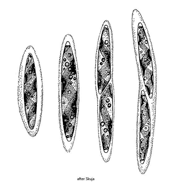
cells spindle-shaped, sometimes cylindrical, apices rounded
length 26–48 µm, width 4–7 µm
with hyaline mucous sheath, 2–5 µm thick
one ribbon-shaped, clockwise spirally chloroplast, 1.5–3 turns
chlorplast with 2–4 pyrenoids
both ends of the chloroplast with orange carotinoid bodies
nucleus central
Spirotaenia erythrocephala
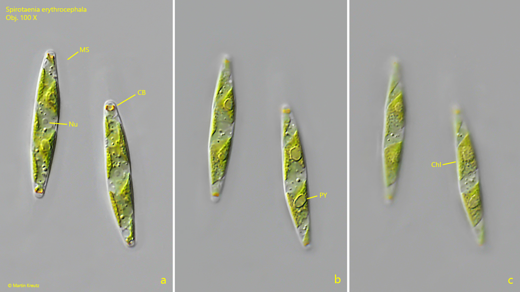
I find Spirotaenia erythrocephala very rarely and exclusively in the Simmelried. This desmid alga can be easily identified by its characteristic orange carotenoid bodies, which are located at the end of the chloroplast and thus in the apices of the cell (s. fig. 1 a). They appear to be ring-shaped, as described by Skuja (1964). The ribbon-shaped choroplast is spiralized in a clockwise direction and lies against the cell wall (s. fig. 1 c). In my specimens mostly 2 pyrenoids were present. The delicate mucous sheath is hard to see in the DIC, but still visible (s. fig. 1 a).
Fig. 1 a-c:Spirotaenia erythrocephala. L = 30–31 µm. Three focal planes of two specimen. Note the orange colored carotinoid bodies (CB) at the end of the spirally, ribbon-shaped chloroplast (Chl). The mucous sheath (MS) covering the cells is hard to see in DIC. Nu = nucleus, PY = pyrenoid. Obj. 100 X