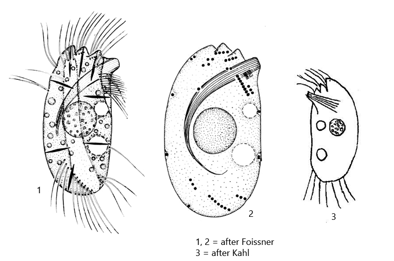
cell oval to almost orthogonal, laterally flattened
pellicle armor-like
left margin straight, right margin convex
three serrated projections at anterior end
three reduced kineties on right side
dorsally and ventrally one kinete each
in the middle of ventral side one bristle-like cilium
deep furrow on right side
length 15–20 µm, width 8–12 µm
macronucleus spherical, located centrally, with small nucleoli
two contractile vacuoles in middle third of body
extrusomes spindle shaped, 3 µm long
cytoplasm clear, with few refractive globules
Stammeridium kahli
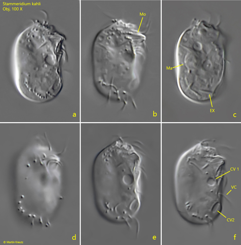
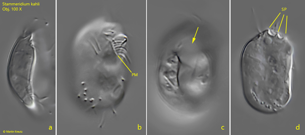
I found this very small ciliate in May 2021 in the Simmelried in the mud layer. At first I thought it was Microthorax, but the typical posterior mouth opening was missing. Instead this is Stammeridium kahli, which was newly described by Foissner in 1985. The anterior mouth opening is snout-like and continued by a basket lined with trichites. I was not able to resolve the trichites of the basket because the images shown below were taken with a dry condenser N.A. 0.9. However, further features are visible, which agree with the description and the drawings of Foissner (see above). Particularly striking, in addition to the snout-like mouth opening, are the 3 serrated projections at the anterior end (s. fig. 2 d), the single ventral cilium (s. fig. 1 f), and the furrow on the right side of the body (s. fig. 2 c). The ciliation of the right side of the body is similar to that of Microthorax, with interrupted rows of cilia. There is a paroral membranelle on the right margin of the mouth opening (s. fig. 2 b), as drawn by Foissner (s. above).
Stammeridium kahli was first described by Wenzel in 1953, but obviously Kahl found it in moss samples before and classified it as a “ciliate similar to Microthorax scutiformis“. Kahl could not examine this ciliate in more detail, so no exact description is available from him. However, his drawing (s. drawing 3, above) agrees in many features with the drawings of Foissner.
Fig. 1 a-f:Stammeridium kahli. L = 18 µm. Different focal planes of a freely swimming specimen from the right side. CV 1, CV2 = contractile vacuoles; EX = extrusomes; Ma = macronucleus; MO = mouth opening; VC = ventral single cilia. Obj. 100 X.
Fig. 2 a-d:Stammeridium kahli. L = 18 µm. A ventral view (a) and three focal planes of the frontal view (b -d). Note the furrow on the right side (arrow, c) and the serrated projections (SP) at the anterior end (d). PM = paroral membranelle. Obj. 100 X.
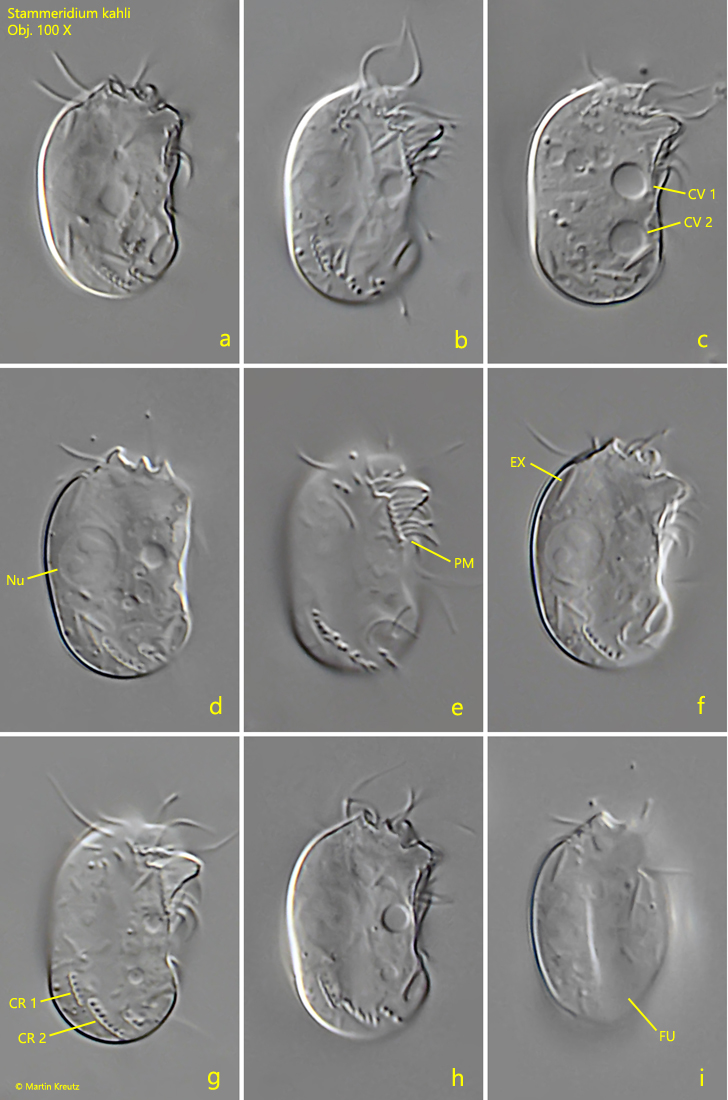
Fig. 3 a-i:Stammeridium kahli. L = 18 µm. Different focal planes from right of a second specimen. CV 1, CV 2 = contractile vacuoles; CR 1, CR 2 = rows of cilia; EX = extrusome; FU = furrow on right side; Ma = macronucleus; PM = paroral membranelle. Obj. 100 X.