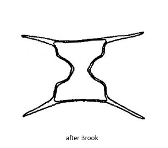
each semi-cell with two spines at the apical edges
apices almost straight, sometimes slightly concave
isthmus about 5–7 µm wide
each semi-cell with a central pyrenoid
nucleus centrally in the isthmus
Staurodesmus extensus
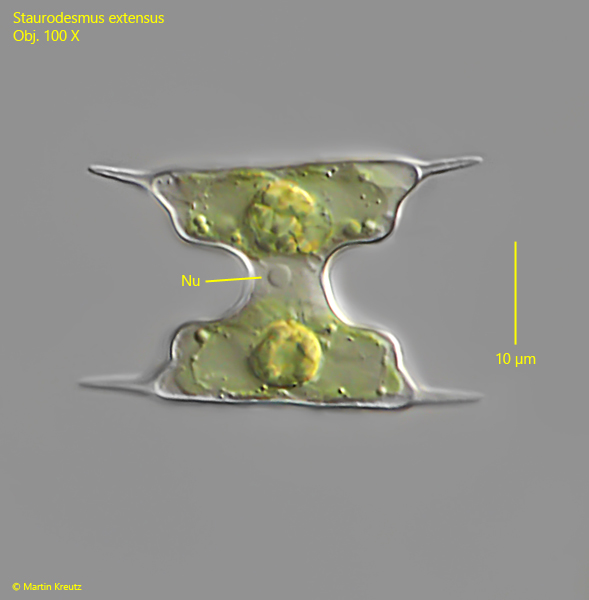
I find Staurodesmus extensus regularly in the Simmelried, both in floating plant masses and in the mud. Although this desmid alga is very small, it can be recognized quite well even at low magnifications by its spines, which protrude almost horizontally from each semi-cell. In addition, the semi-cells taper abruptly to the isthmus, resulting in an almost right-angled transition. Staurodesmus extensus secretes a mucilage sheath, but this can usually only be seen when the sheath is interspersed with fine filaments (s. fig. 3).
Fig. 1:Staurodesmus extensus. L = 21 µm. A slightly squashed specimen with the nucleus (Nu) centrally located in the isthmus. Obj. 100 X.
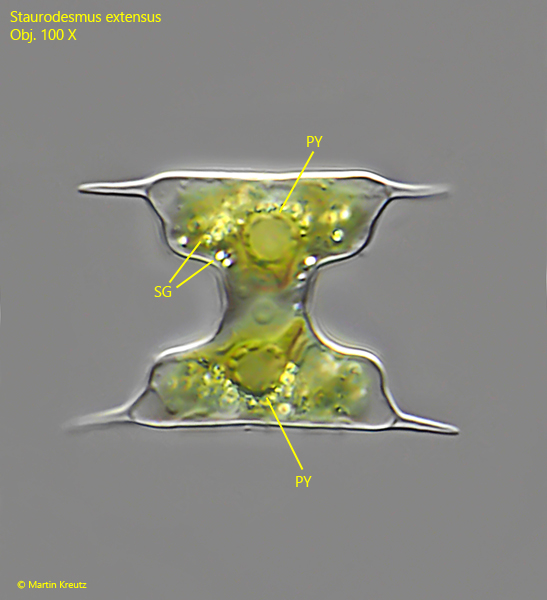
Fig. 2:Staurodesmus extensus. L = 22 µm. A second specimen with the pyrenoids (PY) in each semi-cell surrounded by starch grains (SG), shining bright in DIC. Obj. 100 X.
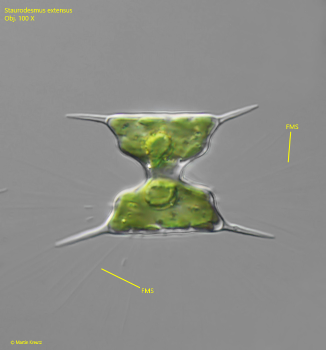
Fig. 3:Staurodesmus extensus. L = 22 µm. This specimen is surrounded by a mucilage sheath. Note the delicate filaments in the mucilage sheath (FMS). Obj. 100 X.