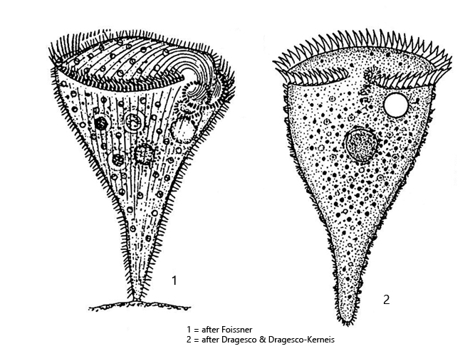
appears violet or brownish by purpel colored cortical granules
spherical symbiotic algae arranged mainly beneath the pellicle
length 250–500 µm (of elongated specimen), contractile by about a third
adoral membranelle running in clockwise direction to oral funnel
macronucleus globular (20–30 µm) in center of body
many (>20) micronuclei surrounded by pigment granules adjacent to the macronucleus
contractile vacuole on left wall of oral funnel
does not build hyaline case
Stentor amethystinus
Stentor amethytinus is by far the most common species of the genus Stentor in the Simmelried area where I found it. In the sample beakers they form sometimes black coatings on the side facing the light. Stentor amethystinus is particularly common in the summer months.
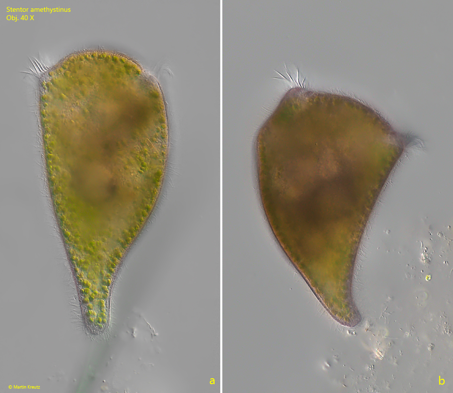
At low magnifications, the specimens usually appear black, as they are opaque due to the symbiotic algae and the purple granules. The specimens rarely settle under the cover glass. Extended specimens are funnel-shaped with a short stalk (s. fig. 1 a-b). This distinguishes them from other Stentor species, which are usually slenderly elongated and trumpet-shaped. Another characteristic feature are the purple-colored granula, which are arranged in rows in the pellicle (s. figs. 2 and 3). The specimens are usually very strongly purple in color. The colored granules are also partially distributed in the cytoplasm. Symbiotic algae are densely arranged below the pellicle. The algae all have their own nucleus, but do not appear to be of the Chlorella type, as they do not have a pyrenoid (s. fig. 7). The spherical macronucleus is surrounded by many micronuclei. According to the literature there are more than 20. In my population there were usually fewer, but more than 10. Each micronucleus is surrounded by a layer of purple granules, which makes them easily recognizable (s. figs. 5 and 6).
Fig. 1 a-b:Stentor amethystinus. Two fully elongated specimens with a length of 232 µm (a) and 200 µm (b). Obj. 40 X.
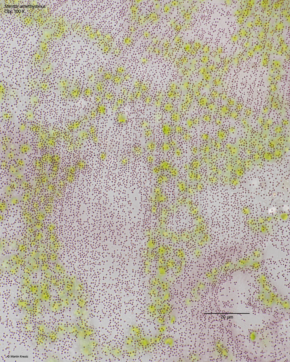
Fig. 2:Stentor amethystinus. The pink colored cortical granules in brightfield illumination. The granules have a diameter of 0.5–0.6 µm. Obj. 100 X.
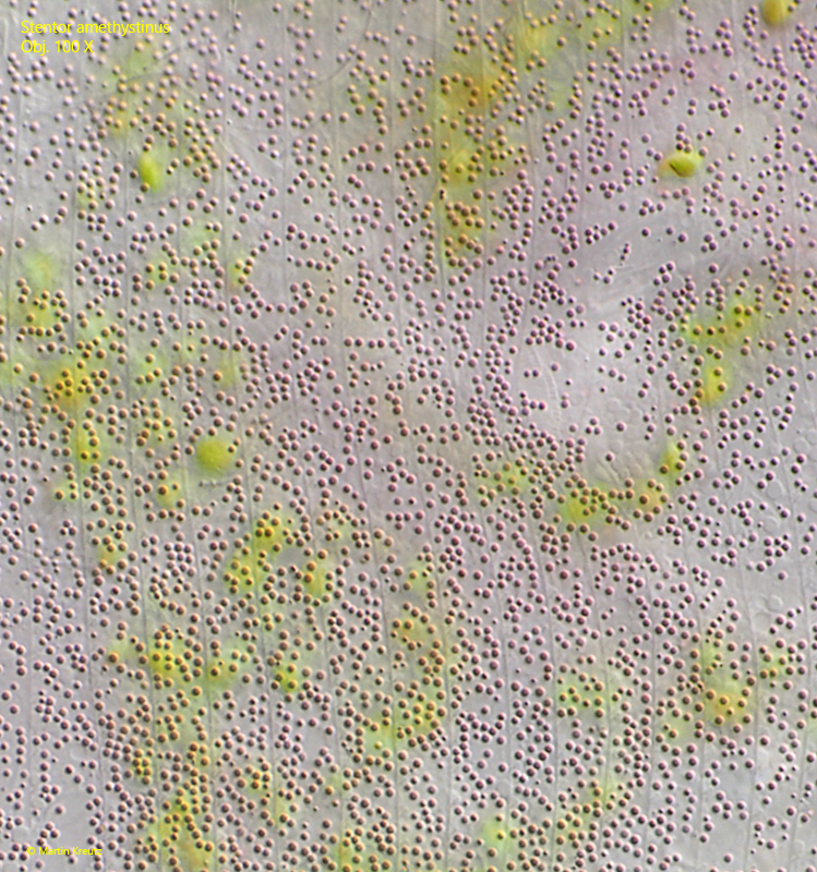
Fig. 3:Stentor amethystinus. The pink cortical granules in DIC. Obj. 100 X.
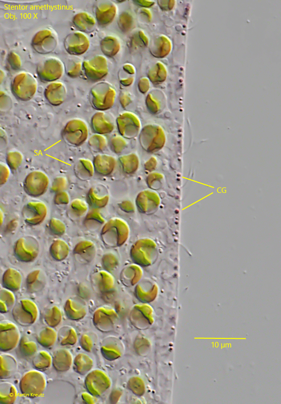
Fig. 4:Stentor amethystinus. The colored cortical granules (CG) are arranged mainly in the pellicle while the symbiotic algae (SA) are arranged in a thick layer below the pellicle. Obj. 100 X.
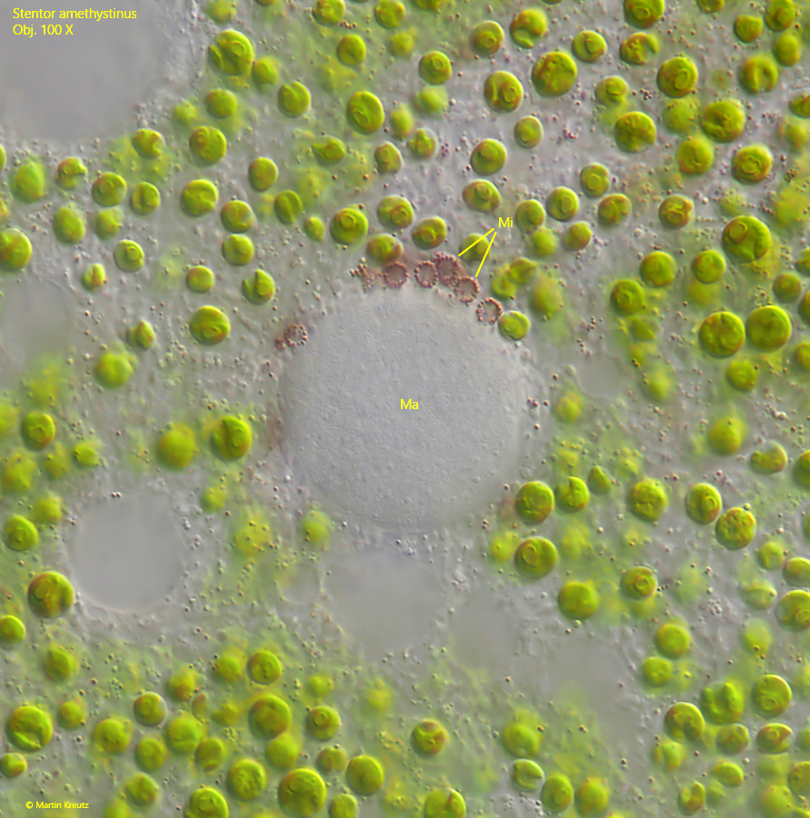
Fig. 5:Stentor amethystinus. The globular macronucleus (Ma) is surrounded by many micronucei (Mi). Each micronucleus is covered with a layer of pink granules. Obj. 100 X.
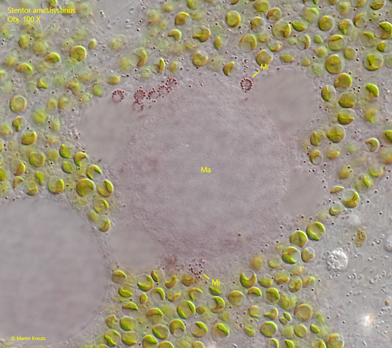
Fig. 6:Stentor amethystinus. The macronucleus (Ma) with the surrounding micronuclei (Mi) in a second specimen. Obj. 100 X.
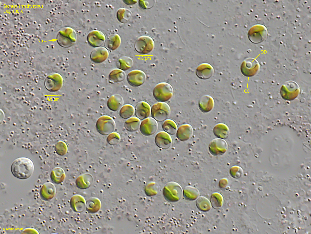
Fig. 7:Stentor amethystinus. The symbiotic algae have a diameter of 4.5–5 µm. The cells have a nucleus (Nu) and a cup-shaped chloroplast (CC). In almost all cells an oily droplet (OD) is present but no pyrenoid. Obj. 100 X.