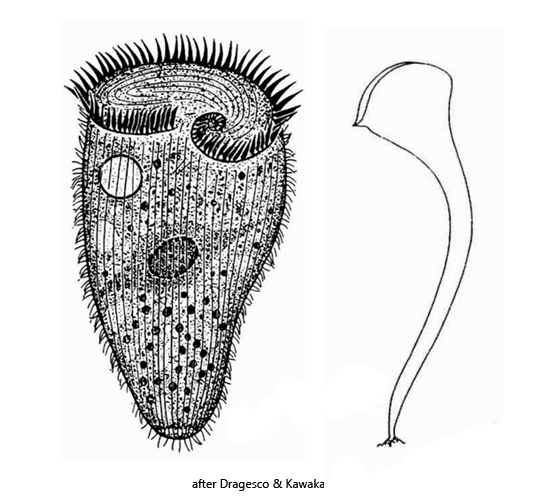
color red-orange, orange-brown or yellow-brown due to pigment granules
in brightfield illumination almost black due to symbiotic algae
anterior third contractile
Stentor fuliginosus
Stentor fuliginosus is very common in the Simmelried. I find this species mainly in spring and early summer. Often I could observe mass developments together with Stentor amethystinus. However, the number of Stentor fuliginosus specimens was always significantly smaller compared to the population of Stentor amethystinus.
Stentor fuliginosus is a species in the genus Stentor that has been insufficiently described. In my population, all specimens were distinctly orange-brown in color. Some specimens were more weakly pigmented, with the green symbiotic algae being more conspicuous. The specimens were almost all about 300 µm long, and some were as long as 370 µm (when elongated). I could always observe only one, spherical macronucleus with 8-10 adjacent micronuclei. The micronuclei were not surrounded by a layer of pigment granules as it is the case in Stentor amethystinus. The pigment granules are not spherical (as in Stentor amthystinus and Stentor coeruleus), but varied in size and shape. They seemed to have with a more crystalline character (s. figs. 5 and 6). I always found the highest concentration of pigment granules in the cytoplasm. In the pellicle the granules were rather sparse and the stripe-like arrangement of the granules was not always recognizable (s. fig. 4). The pigment granules were mostly concentrated around the macronucleus and in the anterior third of the body.
In some individuals I could detect a mucous lorica. However, this was always only indirectly recognizable, by adhering bacteria (s. fig. 3 a-b). I assume that this lorica is built by Stentor fuliginisosus itself, although I could not observe the process. The presence of a lorica has not yet been described for Stentor fuliginosus.
I was able to examine the symbiotic algae of Stentor fuliginosus in detail (s. fig. 6). They are not of the Chlorella type, because they have a pyrenoid and the chloroplast is also not bell-shaped. Furthermore, these algae are larger than Chlorella with a diameter of up to 7.7 µm. I could recognize a cell nucleus at their own in some of these symbiotic algae, but unfortunately I could not document it photographically.
Fig. 1: Stentor fuliginosus. A group of specimens settling on an empty carapace of a water flea. Obj. 10 X.
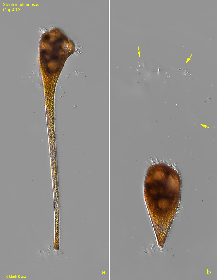
Fig. 2 a-b:Stentor fuliginosus. An elongated specimen (a) with a length of 297 µm and the same specimen contracted (b) with a length of 115 µm. Due to attached bacteria a delicate mucous lorica is visible (arrows). Obj. 40 X.
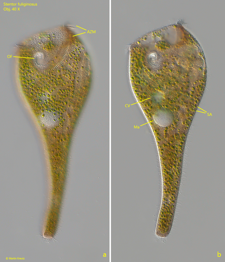
Fig. 3 a-b:Stentor fuliginosus. L = 320 µm. Two focal planes of a slightly squashed specimen. AZM = adoral zone of membranelles, CV = contractile vacuole, Ma = macronucleus, OF = oral funnel, SA = symbiotic algae. Obj. 40 X.
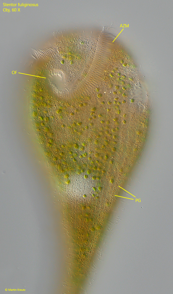
Fig. 4:Stentor fuliginosus. Focal plane on the pellicle of the same specimen shown in fig. 3 a-b. The pigment granules (PG) are of different size and not spherical. Only a low concentration is embedded in the pellicle, so that their stripe-like arrangement is not easy to recognize. The highest concentration of the pigment granules is in the cytoplasm. AZM = adoral zone of membranelles, OF = oral funnel. Obj. 60 X.
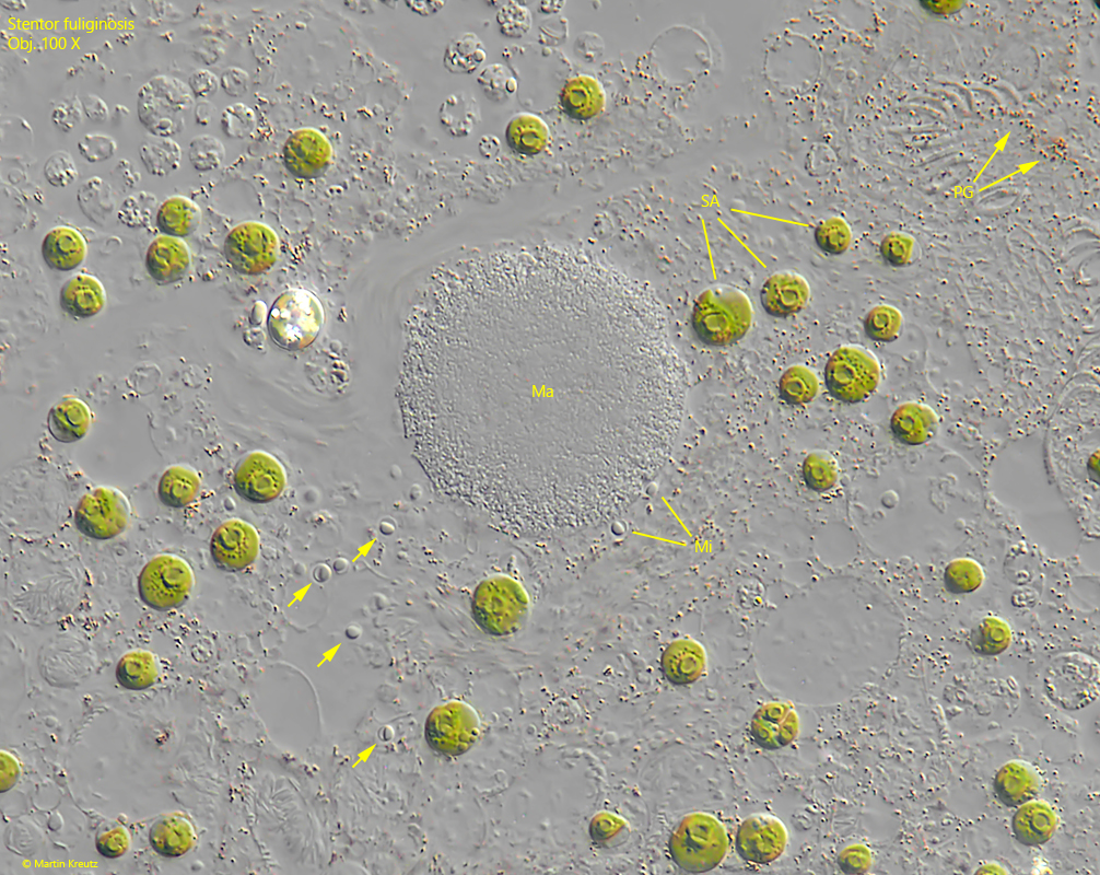
Fig. 5:Stentor fuliginosus. The globular macronucleus (Ma) and the adjacent micronuclei (Mi) in a strongly squashed specimen. Note the micronuclei in the cytoplasm (arrows) detached from the macronucleus due to coverslip pressure. PG = pigement granules in the cytoplasm. Obj. 100 X.
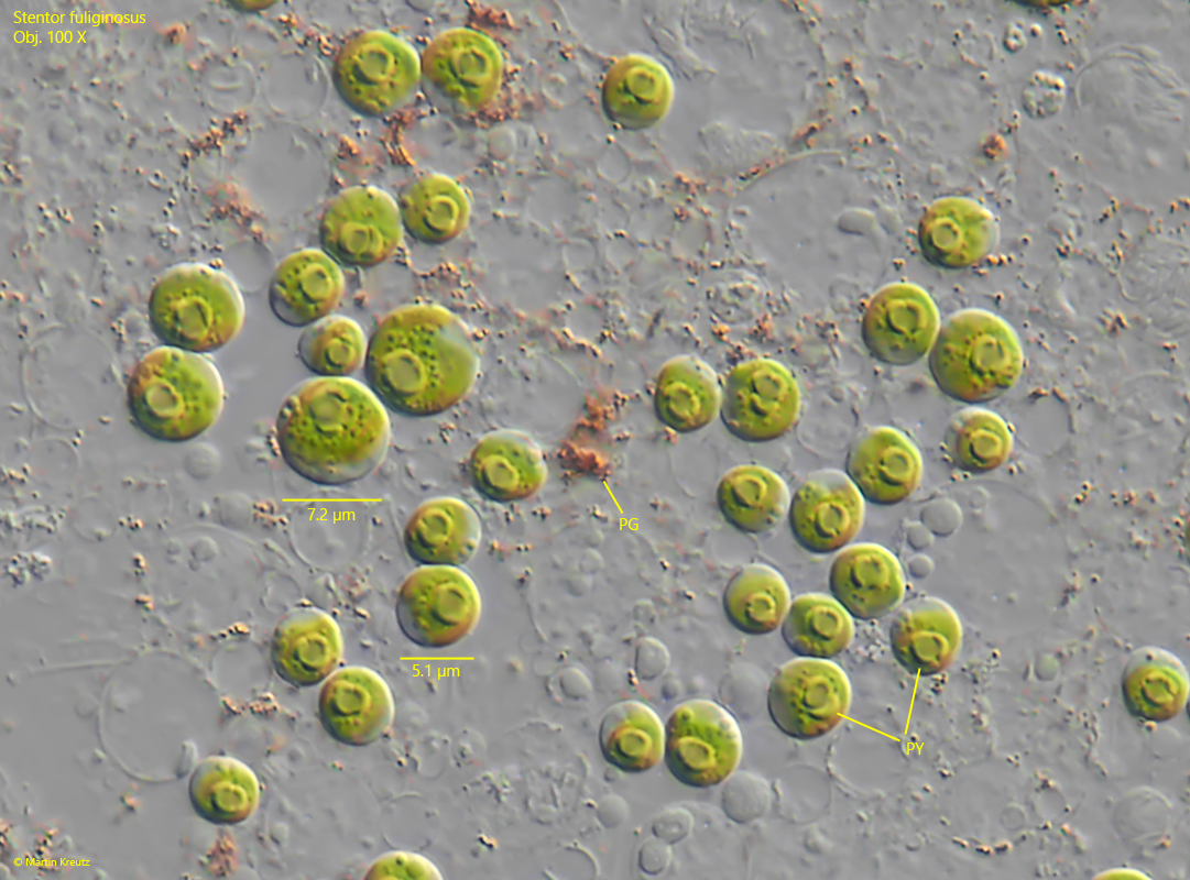
Fig. 6:Stentor fuliginosus. The symbiotic algae in the cytoplasm in detail. The alage are not of the Chlorella type. A distinct pyrenoid with a shell of starch grains is present (in each cell) and the chloroplast is not cup-shaped. The size of the symbiotic algae ist 4.5–7.5 µm. PG = pigment granules in the cytoplasm. Obj. 100 X.