appears blueish to sea-green due to colored cortical granules
no symbiotic algae
length 200–500 µm (of elongated specimen)
adoral membranelle running in clockwise direction to oral funnel
attached to substrate with thigmotactic cilia
macronucleus globular in center of body
many flattened micronuclei adjacent to the macronucleus
contractile vacuole on left wall of oral funnel with short anterior and long posterior collecting duct
presence of hyaline case not confirmed
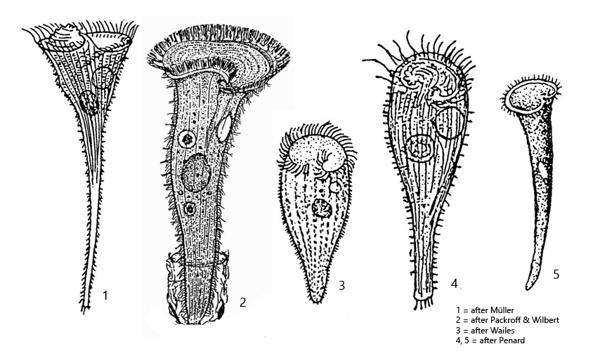
Stentor multiformis
Stentor multiformis is one of the Stentor species that I find comparatively rarely. So far I have only found Stentor multiformis in the mud of the Simmelried.
The main characteristics of Stentor multiformis are a blueish or sea-green coloration and a spherical macronucleus in the middle of the cell. Due to its blueish coloration, it can easily be confused with Stentor coeruleus. However, Stentor coeruleus has a moniliform macronucleus and is also 3-6 times larger than Stentor multiformis.
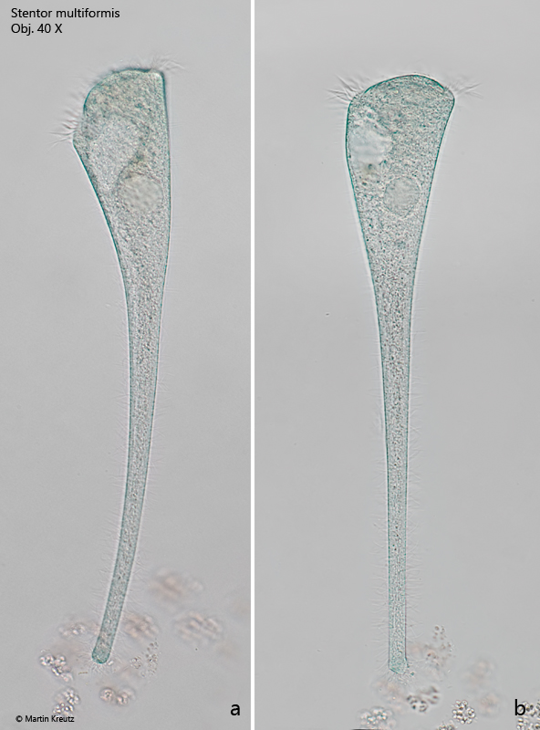
The specimens of my population of Stentor multiformis were mostly sea-green in color, although the intensity of the coloration varied greatly. In addition to intensely colored specimens (s. fig. 1 a-b), I also found almost colorless ones (s. fig. 4 a-b). I could not exactly determine the number of micronuclei attached to the macronucleus, but there were more than 5. The shape of the micronuclei is described as flattened by Foissner et al. (1992). I could not recognize such a flattening in my specimens. Rather, they appeared to be spherical (s. fig. 5). The outstretched specimens of my population were mostly 300–320 µm long.
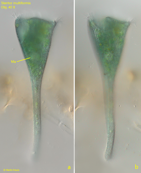
Fig. 1 a-b:Stentor multiformis. L = 360 µm. A fully elongated specimen with an intense sea green color. Obj. 40 X.
Fig. 2 a-b:Stentor multiformis. L = 350 µm. A second elongated specimen with a faint coloration. Obj. 20 X.
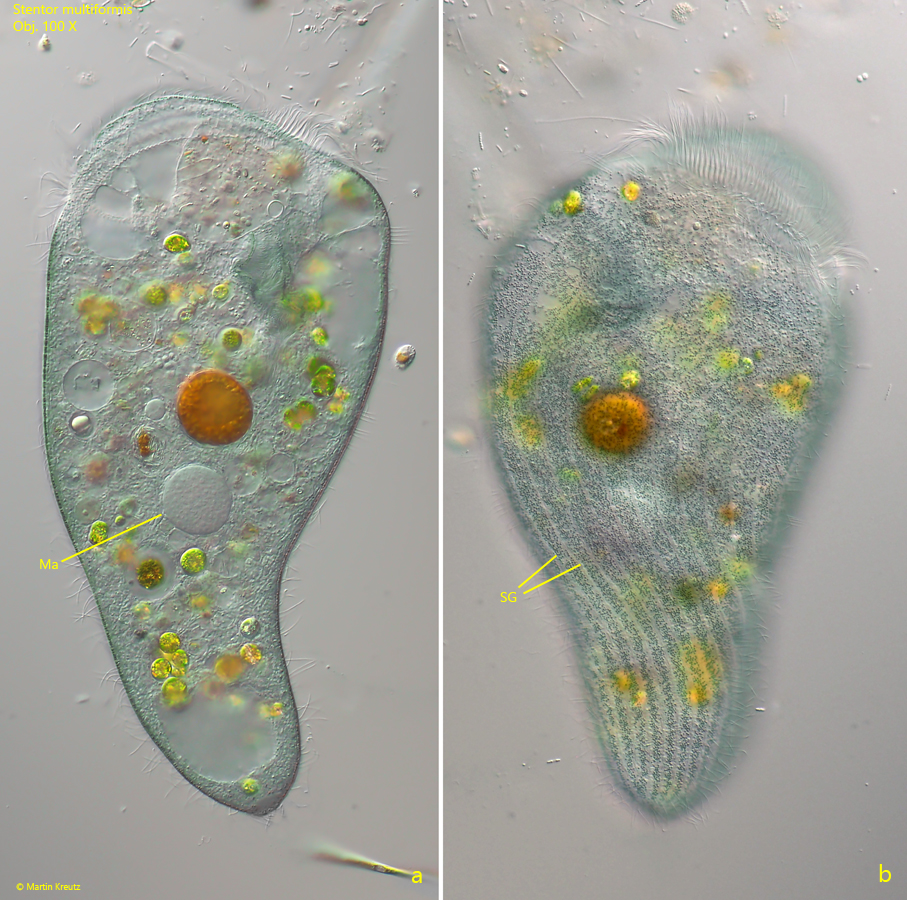
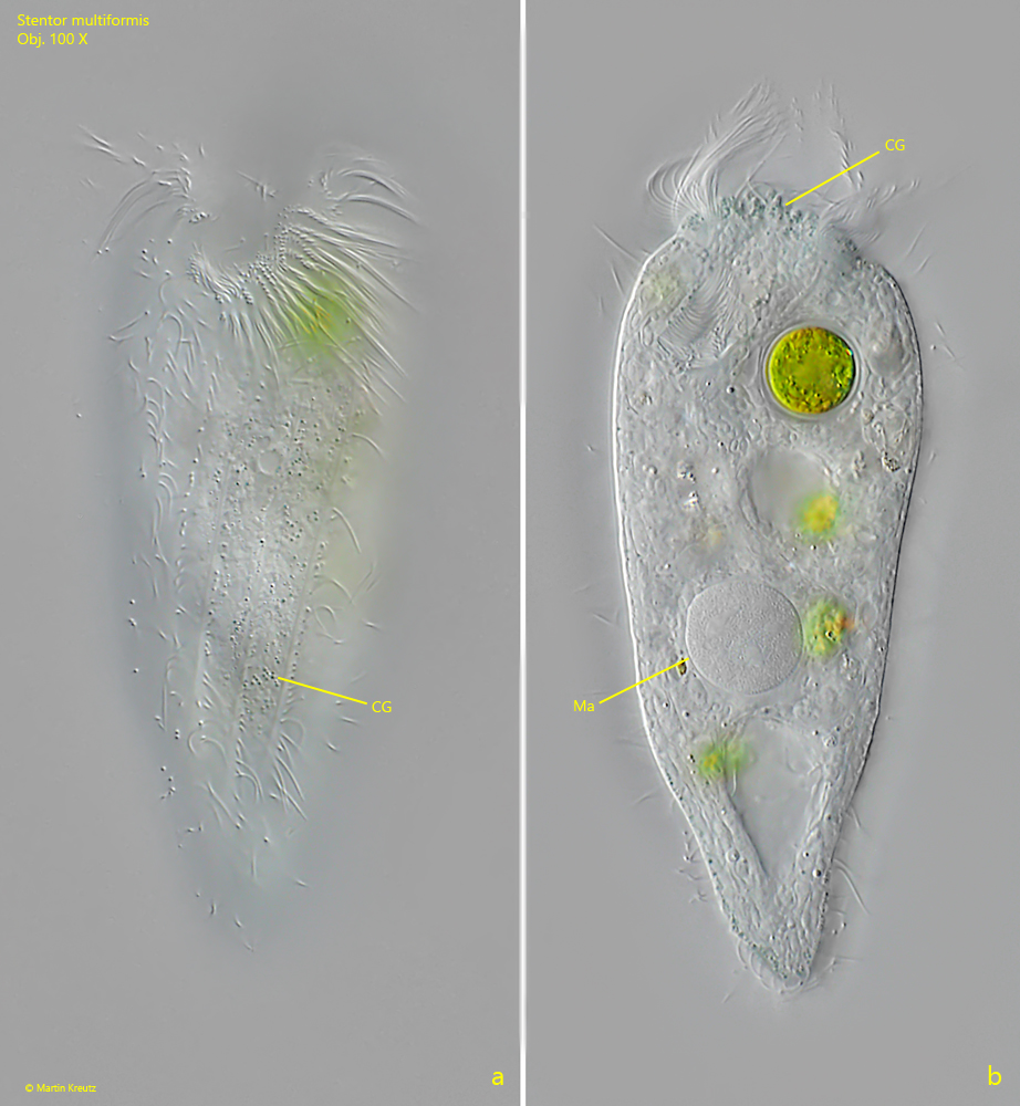
Fig. 3 a-b:Stentor multiformis. Two focal planes of a slightly squashed specimen. Note the sea green coloration and the centrally located spherical macronucleus (Ma). Obj. 100 X.
Fig. 4 a-b:Stentor multiformis. L = 105 µm. A slightly squashed, contracted specimen. This specimen is almost colorless with only a few colored cortical granules (CG). Ma = macronucleus. Obj. 100 X.
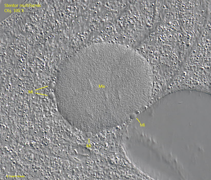
Fig. 5:Stentor multiformis. The spherical macronucleus (Ma) in a strongly squashed specimen with some of the adjacent micronuclei (Mi). Obj. 100 X.