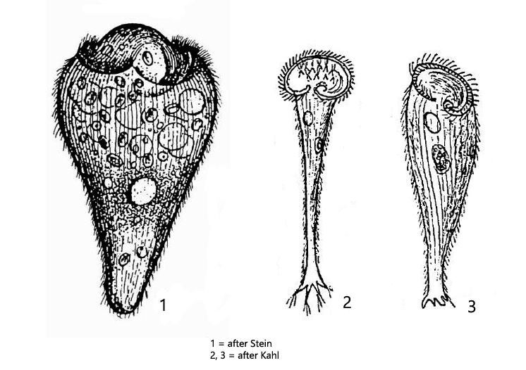
body funnel-spaped, sometimes slightly triangular, contractile
length 200–350 µm
one contractile vacuole on left wall of oral funnel
under pellicle bands of brown-yellowish or brown pigment granules
adorale zone of membranelles leads into mouth opening in a rightward coiled spiral
one spherical or ellipsoid macronucleus in center of cell
no symbiotic algae
Stentor niger
Stentor niger is very rare and there are only very few records. The species was described in 1992 by Foissner, Berger and Kohmann in their “Revision der Ciliaten des Saprobiensystems, volume II” (s. literature). However, the authors stated: “wir haben Stentor niger nie gefunden (we have never found Stentor niger)”.
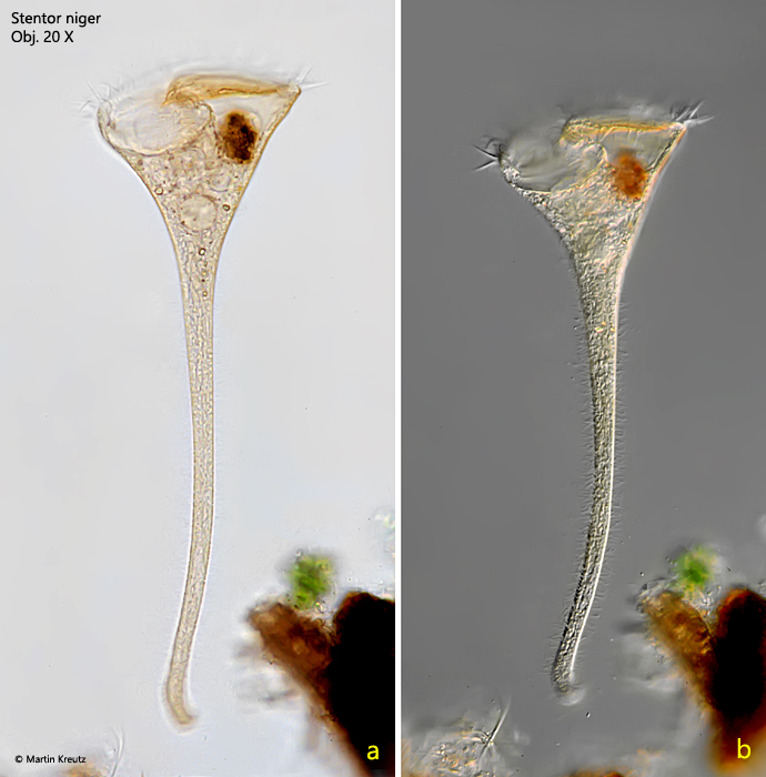
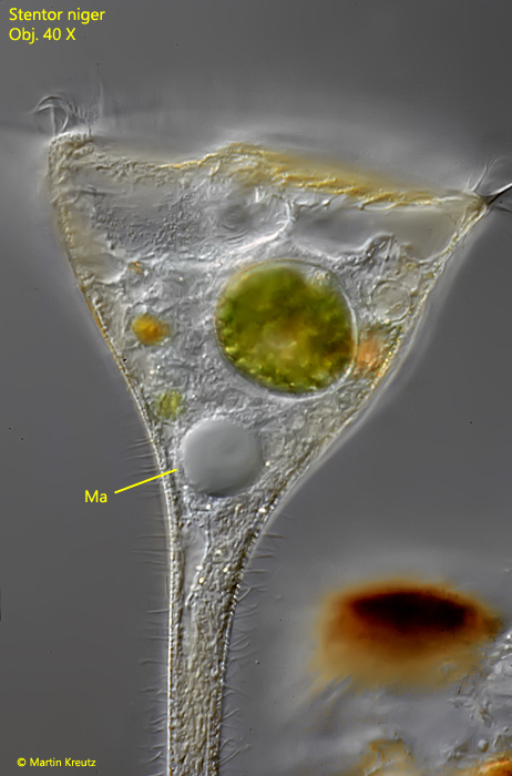
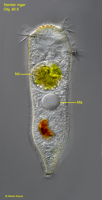
I was able to find few specimens of Stentor niger in a sample from the Edtmoor in Austria in 1998. The images shown below are from these specimens. Six years later I found another 2 specimens of Stentor niger in the Simmelried. After that I have not foud this species again. On the specimens found so far I could verify the description of the earlier authors. As described by Stein and Kahl, I could not detect any symbiotic algae in the specimens, which justifies the separation to Stentor amethystinus. My specimens were yellow-brownish in color, but never brown or even black (s. fig. 1 a-b). Furthermore, my fully extended specimens were up to 436 µm long and thus much longer than indicated by previous authors (200–350 µm). however, the measured length depends strongly on the “degree of extension”. The length of contracted, free-swimming specimens is about 200 µm (s. fig. 3). The other characteristics are consistent with the description of the species. The round macronucleus is located in the middle of the cell (s. figs. 2 and 3). So far, however, no information was available on the number and position of the micronuclei. I could observe 6 spherical micronuclei around the macronucleus in a strongly squashed specimen (s. fig. 4). The pigmented granules are arranged in bands separated by the kineties. In my specimen the granules were yellow-brownish in color (s. fig. 5). Kahl observed that Stentor niger can form root-like projections when attaching to solid substrates (s. drawings 2 and 3 above). I could not observe any such projections in my specimens.
Fig. 1 a-b:Stentor niger. L = 436 µm. A fully extended specimen in brightfield illumination (a) and DIC (b). Note the yellow-brownish color of the specimen. Obj. 20 X.
Fig. 2:Stentor niger. Focal plane on the spherical macronucleus in mid-body of the fully extended specimen shown in fig. 1 a-b. Obj. 40 X.
Fig. 3:Stentor niger. L = 207 µm. A contracted, freely swimming specimen. Obj. 40 X.
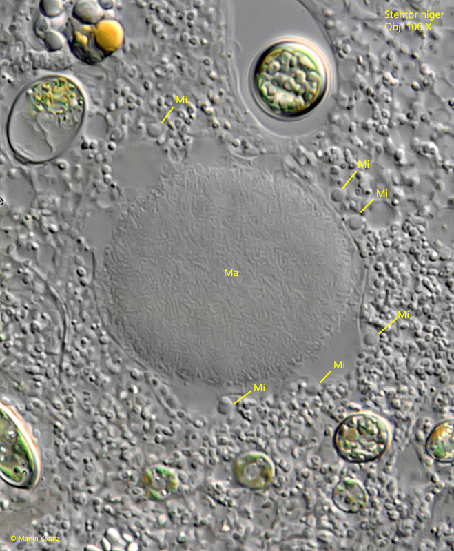
Fig. 4:Stentor niger. The spherical macronucleus (Ma) and 6 adjacent micronuclei (Mi) in a strongly squashed specimen. Obj. 100 X.
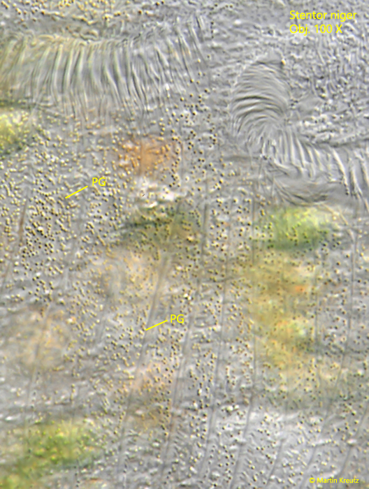
Fig. 5:Stentor niger. The brown yellowish pigment granules (PG) beneath the pellicle are arranged in bands, separated by the kineties of cilia. Obj. 100 X.