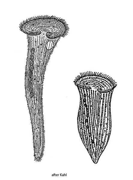
body elongated trumpet-shaped, contracted specimens obovoid to club-shaped
length up to 2000 µm (of elongated specimens)
cytoplasm green due to symbiotic algae (Chlorella)
adoral membranelle running in clockwise direction to oral funnel
attached to the substrate with thigmotactic cilia
macronucleus moniliform, 5–20 nodules
contractile vacuole on left wall of oral funnel
Stentor polymorphus
Stentor polymorphus is a very common ciliate that I find regularly in most of my sampling location. In July 2023, I even observed a mass development of Stentor polymorphus in the Purren pond. Billions of specimens covered the top layer of leaves on the mud, turning them an intense green.
Due to its size and green coloration caused by thousands of symbiotic Chlorella algae, Stentor polymorphus is unmistakable. The macronucleus is monoliform and consists of 5 – 20 nodules. I was able to identify several microsnuclei adjacent to the nodules, which was not previously described by Kahl (1932) or Foissner (1992).
Tactile bristles were observed in Stentor polymorphus by Penard (1922). However, this observation has not been confirmed so far. Also I could not recognize any in my population.
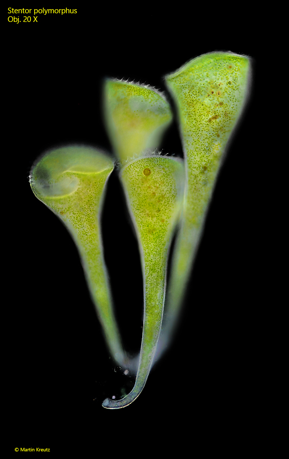
Fig. 1: Stentor polymorphus. L = 550 – 710 µm. A group of four fully extended specimens in darkfield illumination. Obj. 20 X.
Fig. 2: Stentor polymorphus. L = 510 µm. A partly extended specimen. Obj. 40 X.
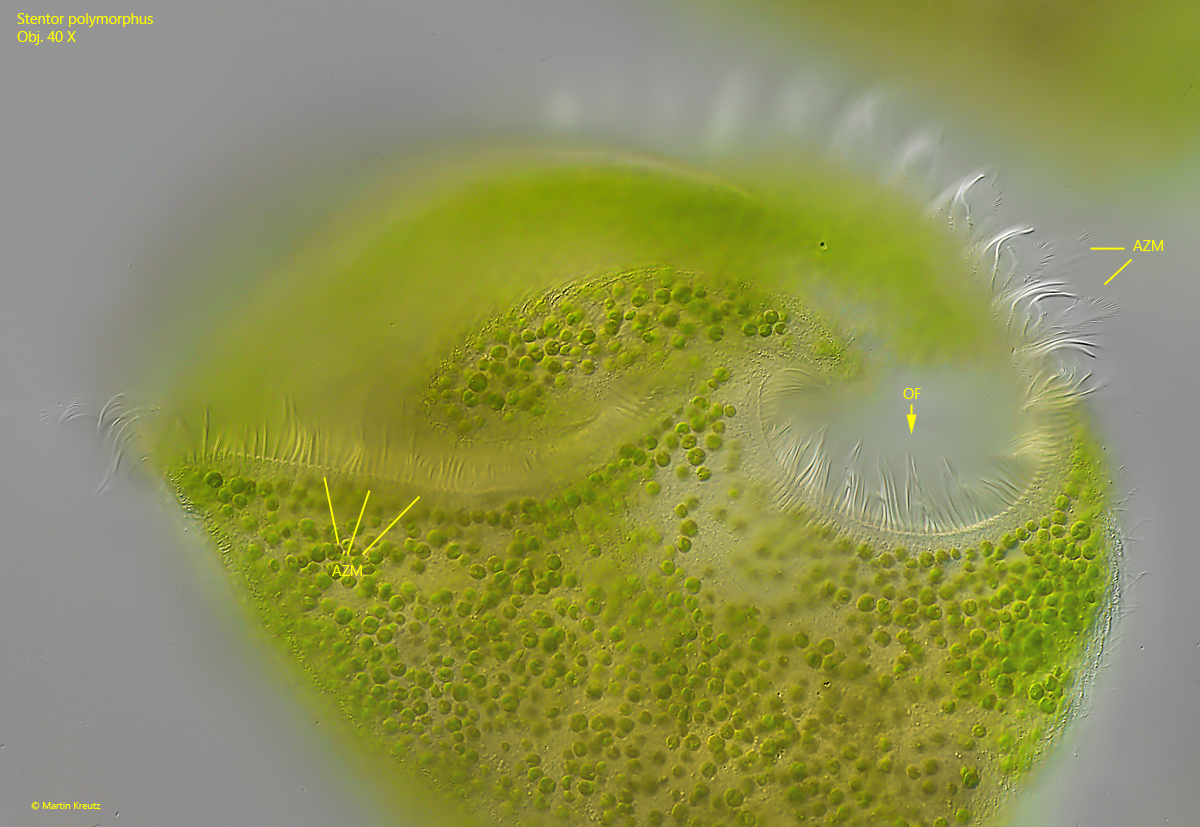
Fig. 3: Stentor polymorphus. The adoral zone of membranelles (AZM) in detail. In apical view the adoral zone is almost bretzel-shaped and runs in clockwise direction into the oral funnel (OF). Obj. 40 X.
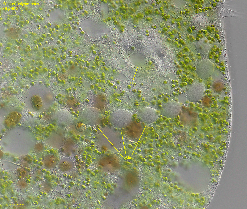
Fig. 4: Stentor polymorphus. Focal plane on the nodules of the moniliform macronucleus (Ma) in a slightly squashed specimen. OF = oral funnel. Obj. 40 X.
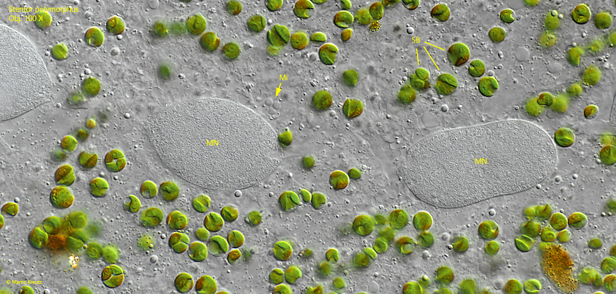
Fig. 5: Stentor polymorphus. The nodules of the macronucleus (MN) in detail. Note the group of 3 micronuclei (Mi) adjacent to the nodule. SB = symbiotic algae. Obj. 100 X.
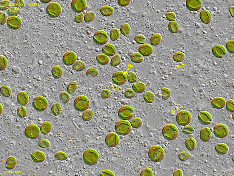
Fig. 6: Stentor polymorphus. The symbiotic algae have a diameter of 4–6 µm and are member of the genus Chlorella. Each Chlorella cell has a nucleus (Nu) of its own which is hard to recognize. Obj. 100 X.