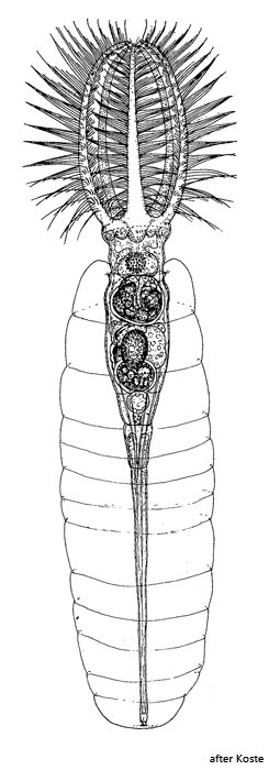
rotifer with corona of 5 tentacles in a gelatinous tube
length up to 1500 µm
length of corona tentacles about one third of body length
tentacles bent to the longitudinal axis form an egg-shaped basket
gelatinous tube about two thirds of body length, with slight constrictions
corona tentacles with long cilia, arranged in two parallel rows
coronal margin between tentacles is naked
wide mouth cavity, large proventriculum
foot one third of body length, with cement glands
eyespots absent
Stephanoceros fimbriatus
I find Stephanoceros fimbriatus rarely in the Simmelried and in the Mühlweiher Litzelstetten but very often in the pond of the convent Hegne. There I find the specimens exclusively on the pinnate leaves of the water hose (Utricularia vulgaris) and the water milfoil (Myriophyllum spec.). The specimens are easily recognized and identified by their size and unique shape.
The 5 tentacles with long cilia form a kind of catching basket. To catch prey, the tentacles are spread so wide that prey either actively swims into them or they are driven into the catch basket on contact with the outer long cilia. Upon contact with a prey organism, the cilia perform a whip-like movement. Once the prey is inside the catch basket, the tentacles close and the prey is directed into the vestibule and from there into the proventriculum with the aid of shorter cilia located on the inside of the tentacles. At the bottom of the proventriculum the trophi are located, which grab the collected prey organisms one by one and crush them to transport them into the stomach below (s. fig. 4 a). Prey organisms are algae, ciliates, small rotifers and even small gastrotrichs.
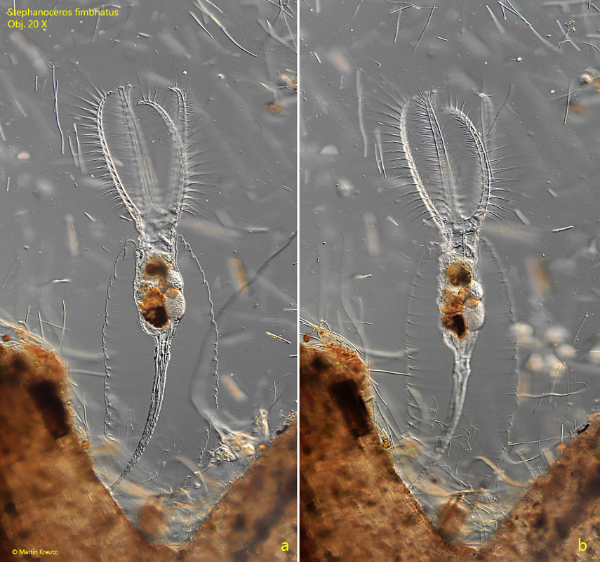
Fig. 1 a-b:Stephanoceros fimbriatus. L = 648 µm. Two focal planes of a fully extended specimen. Obj. 20 X.
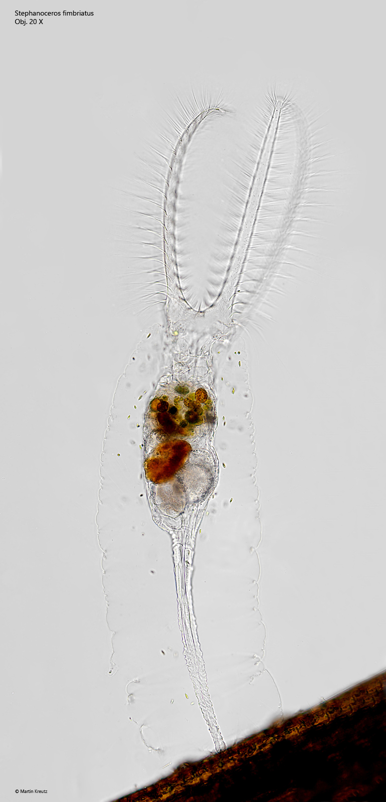
Fig. 2:Stephanoceros fimbriatus. L = 1188 µm. A fully extended specimen in brightfield illumination. Obj. 20 X..
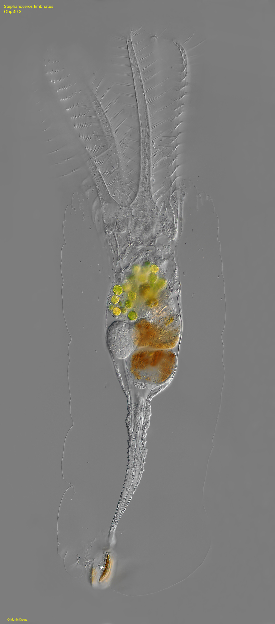
Fig. 3:Stephanoceros fimbriatus. L = 648 µm. A specimen detached from the subtrate. Obj. 40 X.
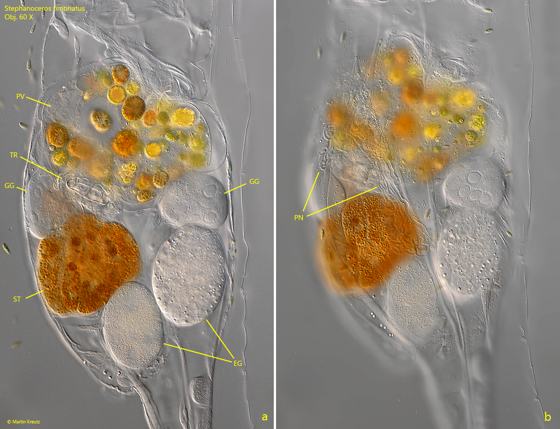
Fig. 4 a-b:Stephanoceros fimbriatus. Two focal planes of the mid-body. EG = eggs, GG = gastric glands, PN = tubes of the protonephridium, PV = proventriculum, ST = stomach TR = trophi. Obj. 60 X.
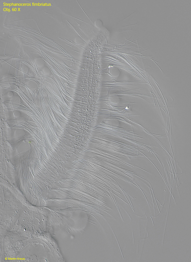
Fig. 5:Stephanoceros fimbriatus. The ciliation of one of the tentacles in a squashed specimen. Obj. 60 X.
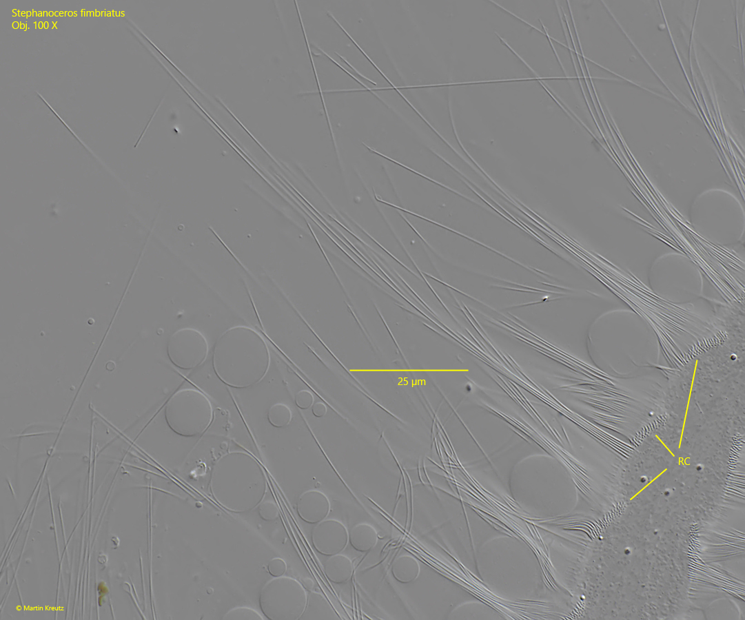
Fig. 6:Stephanoceros fimbriatus. The cilia on the outside of a tentacle in detail. The longest cilia are about 70 µm long. The cilia are arranged in short rows (RC), which run perpendicular to the longitudinal axis of the tentacles. Obj. 100 X.
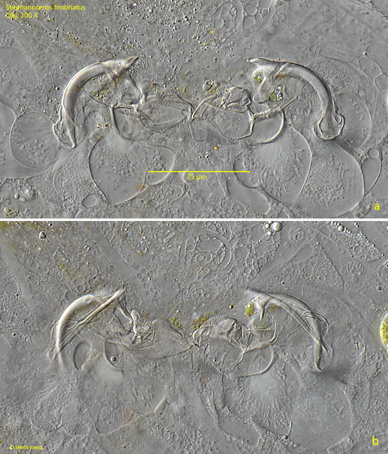
Fig. 7 a-b:Stephanoceros fimbriatus. Two focal planes of the trophi. Obj. 100 X.