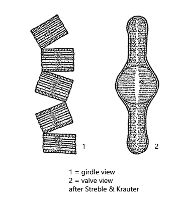
cells are connected at their corners to form zig-zag chains
septa penetrating up to mid-region
in valve view elongated with an inflation in the center
chloroplasts golden brown, lying between the septa
Tabellaria flocculosa
I have found Tabellaria flocculosa so far only in the Ibmer Moor in Austria and in the Simmelried. In the Simmelried this diatom could only be found until 1999, because the moor character of the area has been decreasing more and more. Light microscopically mainly the rectangular or square girdle view is presented. The valve view is only visible when the tabular-shaped diatoms settle on one narrow side, which is virtually never the case when viewed under the coverslip. The cells are connected to each other via their corners to form zig-zag chains, being held together by a gelatinous mass, which is also easily seen by light microscopy (s. fig. 2 a).
Fig. 1 a-b:Tabellaria flocculosa. L = 18–24 µm (of cells). Two focal planes of a zig-zag chain. Obj. 60 X.
Fig. 2 a-b:Tabellaria flocculosa. L = 18–24 µm. Four cells from a zig-zag shaped chain. The cells are connected at the corners via a gelatinous mass (GM). The cell volume is subdivided by septa (SE) reaching up to the mid-region. Between the septa the greenish and golden-brown chloroplasts are arranged. Nu = nucleus. Obj. 100 X.