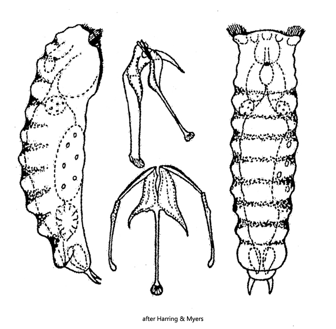
foot rudimentary, toes short, conical and slightly curved
no bumps between the toes
wheel organ of Notommata-type
auricles retractable
retrocerebral organ with attached eyespot
Taphrocampa annulosa
So far I have found only a few specimens of Taphrocampa annulosa. All findings are from the Simmelried. Mostly I discover the specimens when they glide along floating coverslips on their ventral side. The genus Taphrocampa is easily recognized by the folded dorsal epidermis. To distinguish the two similar species Taphrocampa annulosa and Taphrocampa selenura, the shape of the toes must be considered. In Taphrocampa annulosa, the toes are short, curved inward, and taper to a blunt tip. In Taphrocampa selenura the toes are much longer, saber-shaped, and curved inward to a sharp point. In my specimens, the toes were short and blunt, so Taphrocampa annulosa is present here.
The dorsal epidermis of Taphrocampa annulosa is described to be sticky, so adherent foreign particles and bacteria are often found on it. With my (few) specimens I could not observe this.
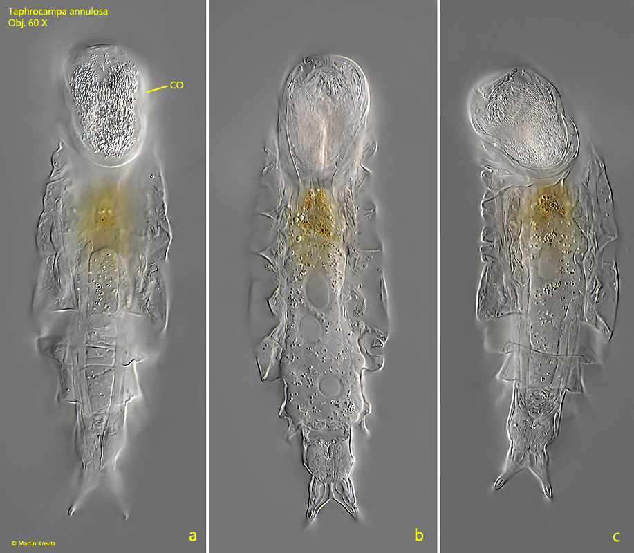
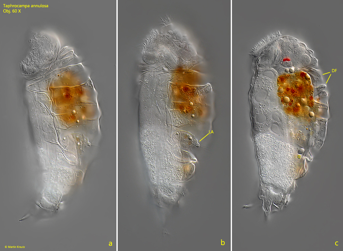
Fig. 1 a-c:Taphrocampa annulosa. L = 178 µm. Ventral view of a freely gliding specimen. Obj. 60 X.
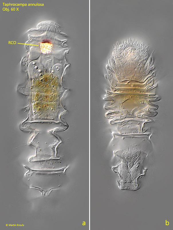
Fig. 2 a-b:Taphrocampa annulosa. L = 178 µm. Dorsal view of the same specimen shown in fig. 1 a-c. Note typical transversal folds especially in the contracted specimen (b). The retrocerebral organ (RCO) shining brightly in DIC. Obj. 60 X.
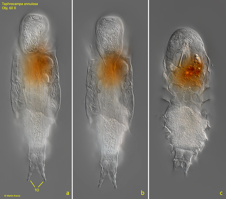
Fig. 3 a-c:Taphrocampa annulosa. L = 201 µm. A second specimen in ventral view. TO = toes. Obj. 60 X.
Fig. 4 a-c:Taphrocampa annulosa. L = 201 µm. Lateral view from left of the same specimen shown in fig. 3 a-c. Note the lateral antenna (LA) and the dorsal folds (DF) of the epidermis. Obj. 60 X.
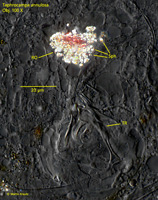
Fig. 5:Taphrocampa annulosa. The retrocerebral organ (RO) in a strongly squashed specimen. It consists of many birefringent spherules (Sph). TR = trophi. Obj. 100 X.
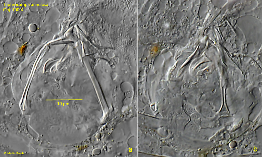
Fig. 6 a-b:Taphrocampa annulosa. Two focal planes of the trophi in a strongly squashed specimen. TR = trophi. Obj. 100 X.