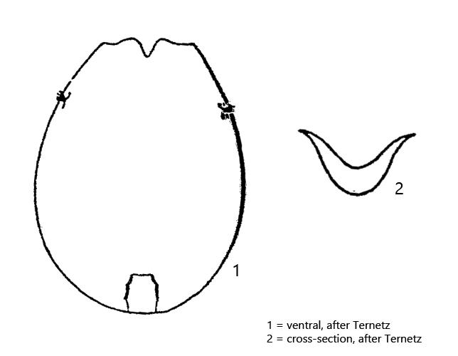
lorica elliptical or ovoid, dorso-ventral flattened
lorica in cross-section trough shaped, with two “wings”
lorica transparent, length 100–110 µm
anterior end of the lorica with V-shaped notch on ventral side
foot opening ventral, at posterior end of lorica, almost rectangular
foot annulated, retractile, distal end with cilia
corona is a band of cilia
two conspicuous retractor muscles
two eyespots
Testudinella incisa
I have found Testudinella incisa only 3 times since 1993. The last finding was in March 2016. All findings are from the Simmelried. In my other finding areas I have not been able to detect the species so far.
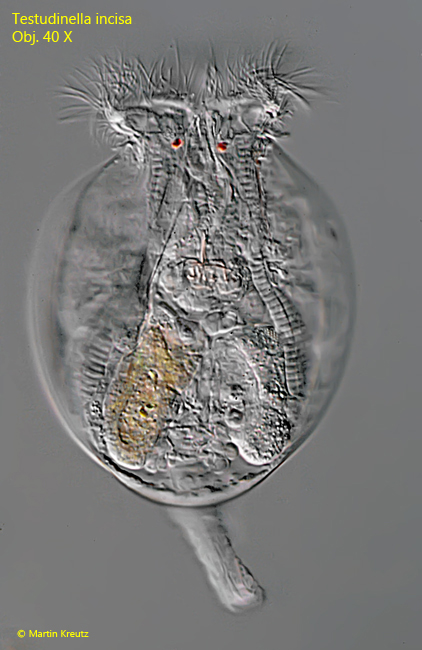
Fig. 1:Testudinella incisa. L = 110 µm (the lorica). Dorsal view of a freely swimming specimen found in September 1996. Because of the trough shape of the lorica, the “wings” are not in the plane of focus. Obj. 40 X.
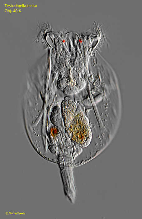
Fig. 2:Testudinella incisa. L = 110 µm (the lorica). Dorsal view of a second freely swimming specimen found in March 2016. Obj. 40 X.
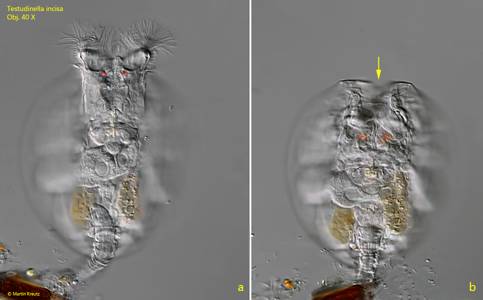
Fig. 3:Testudinella incisa. Ventral view of a fully extended (a) and contracted specimen (b) found in November 2004. The specimen is attached to a detritus flake with the foot. At the anterior end of the lorica a notch is visible (arrow). Obj. 40 X.
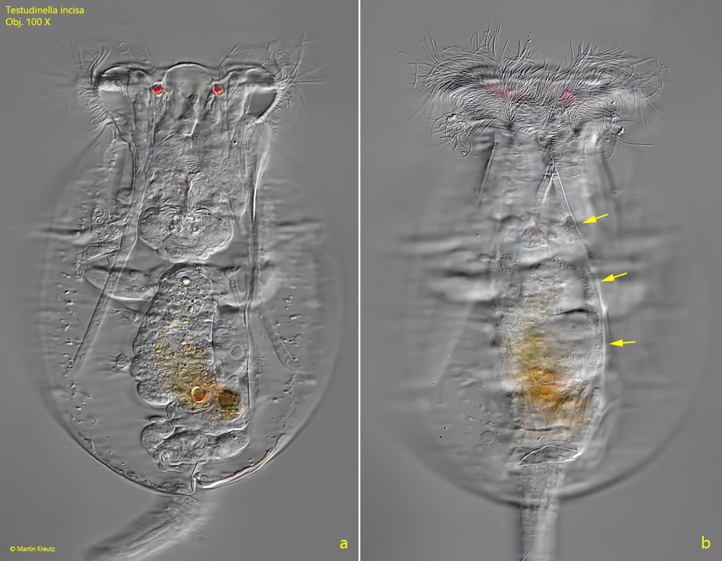
Fig. 4:Testudinella incisa. L = 115 µm (the lorica). Dorsal view of an extended, slightly squashed specimen. The focus on the smooth surface of the lorica (b) shows a wrinkle (arrows), which was created by the trough shape of the lorica under coverslip pressure. Obj. 100 X.