body transparent, length 120–350 µm, width 100–236 µm
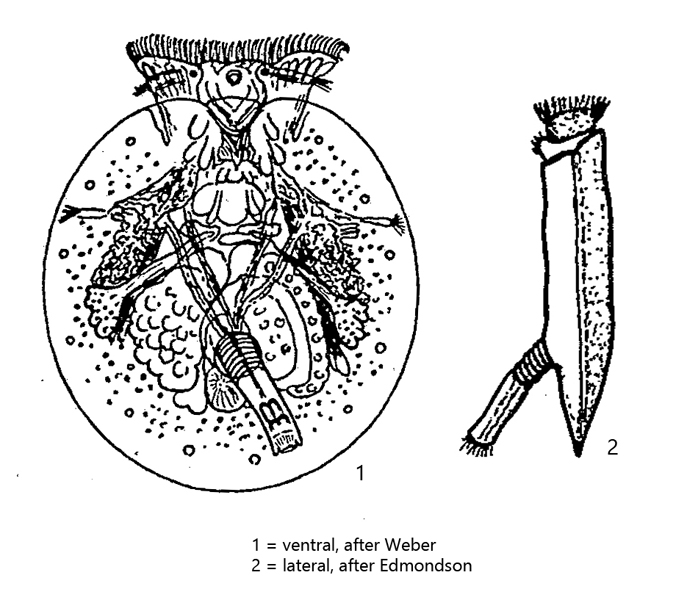
foot opening ventral, circular, almost in the middle
foot annulated, retractile, distal end with cilia
corona is a band of cilia
two conspicuous retractor muscles
two eyespots
Testudinella patina
Testudinella patina is a very common and widespread species. I was able to detect it in several of my sites in floating plant material. The species is very photogenic because of the strongly flattened and transparent lorica. Already in 1995 I took the first photos of Testudinella patina (s. fig. 1) and after that always in larger intervals. I have always been able to find the species in the summer months. Because of the dorso-ventral flattening the internal organs are not on top of each other and one can study the anatomy of this rotifer in detail (s. fig. 4).
Fig. 1:Testudinella patina. L = 200 µm. Ventral view of a slightly squashed specimen. Obj. 60 X.
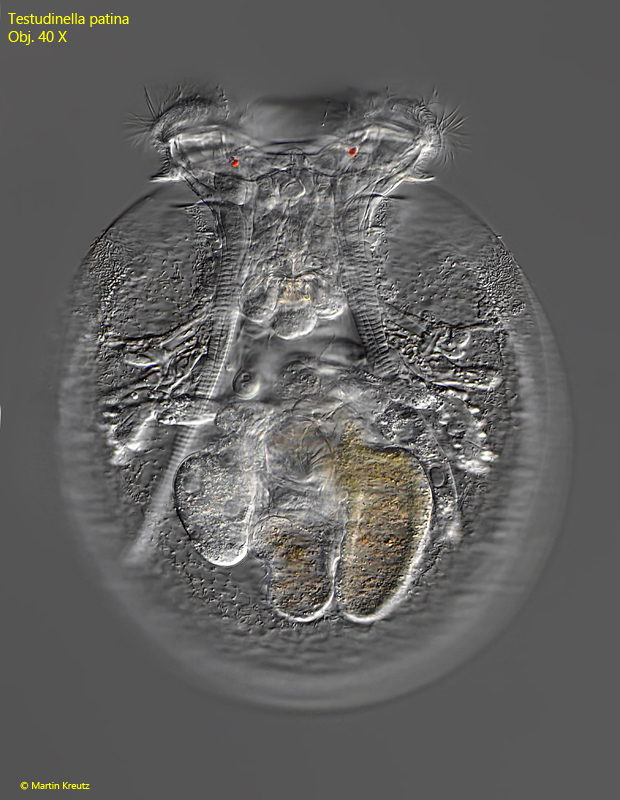
Fig. 2 a-b:Testudinella patina. L = 183 µm. Two focal planes of a ventral view of a slightly squashed specimen. This specimen was found in August 2008. Note the circular foot opening (FO) on the ventral side of the lorica. Obj. 40 X.
Fig. 3:Testudinella patina. L = 256 µm. Ventral view of a slightly squashed specimen. This image was taken in September 2016. Obj. 40 X.
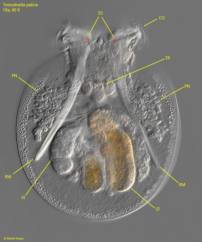
Fig. 4:Testudinella patina. L = 226 µm. Ventral view of a slightly squashed specimen. The image is from Juli 2020. CO = corona, ES = eyespots, PN = protonephridium, RM = retractor mucles, ST = stomach, TR = trophi, VI = vitellarium. Obj. 60 X.