So far, I have only found a single specimen of Thecochaos album in March 2017 in a sample from the Simmelried. It was a Petri dish that I had prepared for floating coverslips. After two weeks, a yellow-green accumulation of Oscillatoria chlorina had formed on the side of the Petri dish facing the light. I examined this cluster of filamentous cyanobacteria and found the specimen of Thecochaos album in it. At low magnification it was similiar to Thecamoeba because of the highly refractive cytoplasm. However, the specimen was much too large for this, which is why I examined it more closely.
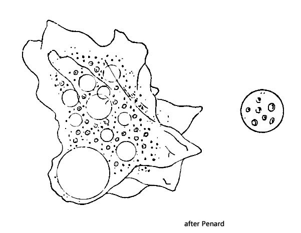
Thecochaos album was first described by Greeff (1891) as Amoeba alba. Penard then found further specimens in 1902 and described them in more detail. He also deposited permanent specimens in the Natural History Museum in London and made a drawing. No further specimens of Thecochaos album were apparently found after 1902. Eighty years later, Page was also unable to find a living specimen, which is why he examined Penard’s permanent specimens in detail in 1981. On the basis of the morphological features found and Penard’s description, he placed the species in the genus Thecochaos under the name Thecochaos album.
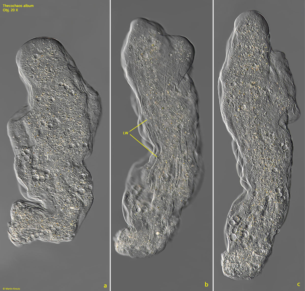
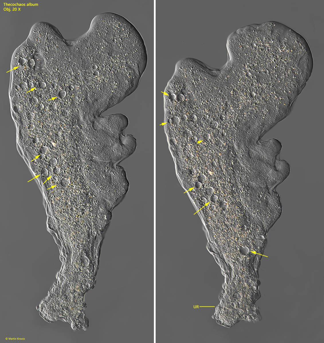
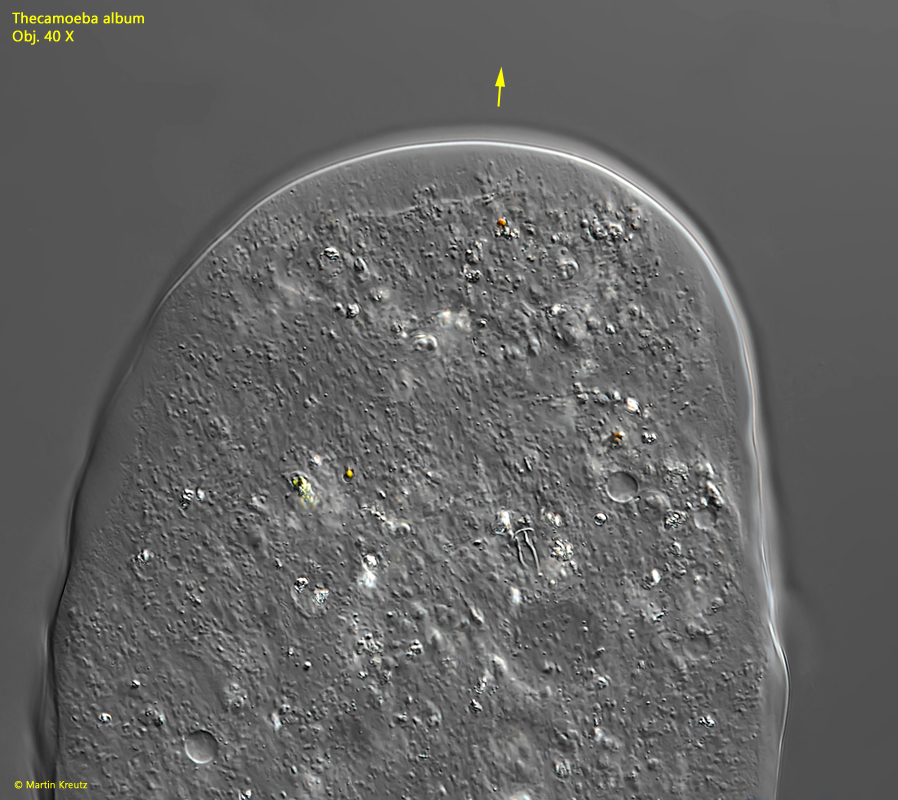
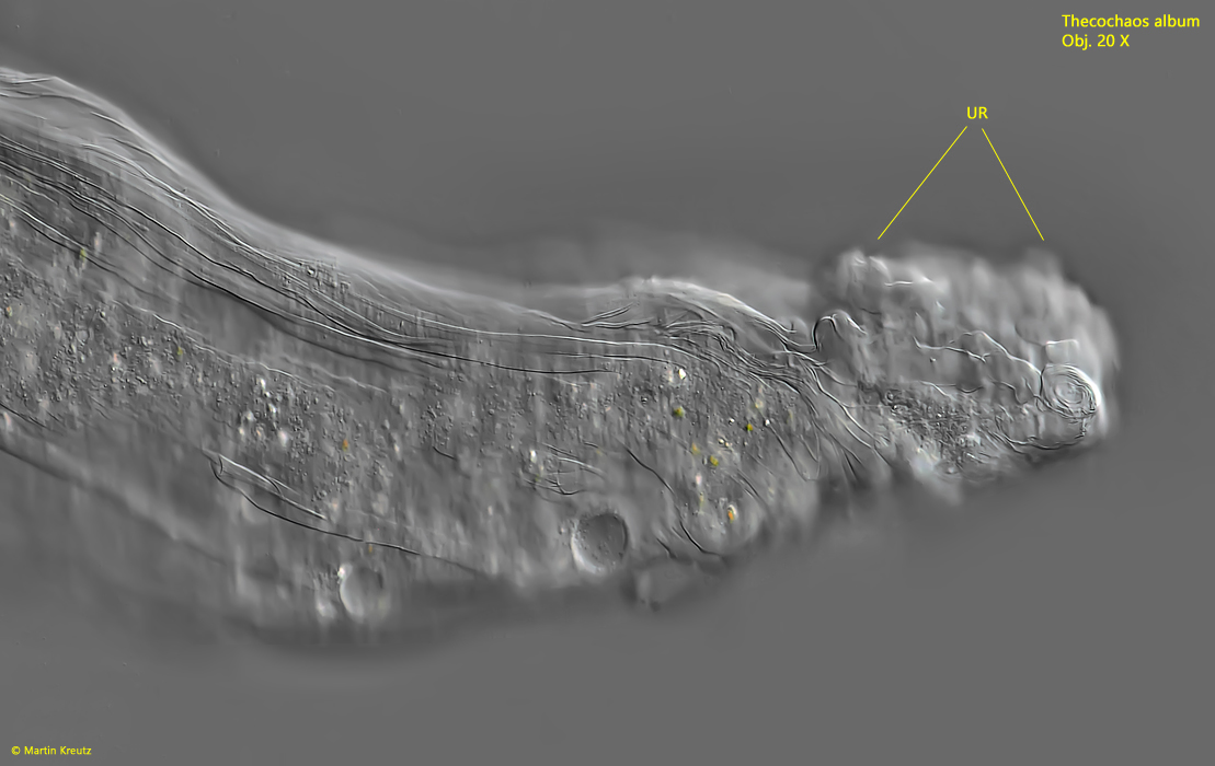
The locomotion flow form of my specimen was mainly monopodial (s. fig. 1 a-c). During locomotion a bulbous uroid is formed (s. fig. 7). The specimen reached a maximum length of 980 µm. Penard gives a maximum size of 360 µm, but for resting specimens. During locomotion, “rivers” of low-viscosity cytoplasm formed in the amoeba, which move very quickly in the flow direction of the amoeba. As this effect is hard to describe, I recorded a short video of it:
Locomotion Thecochaos album
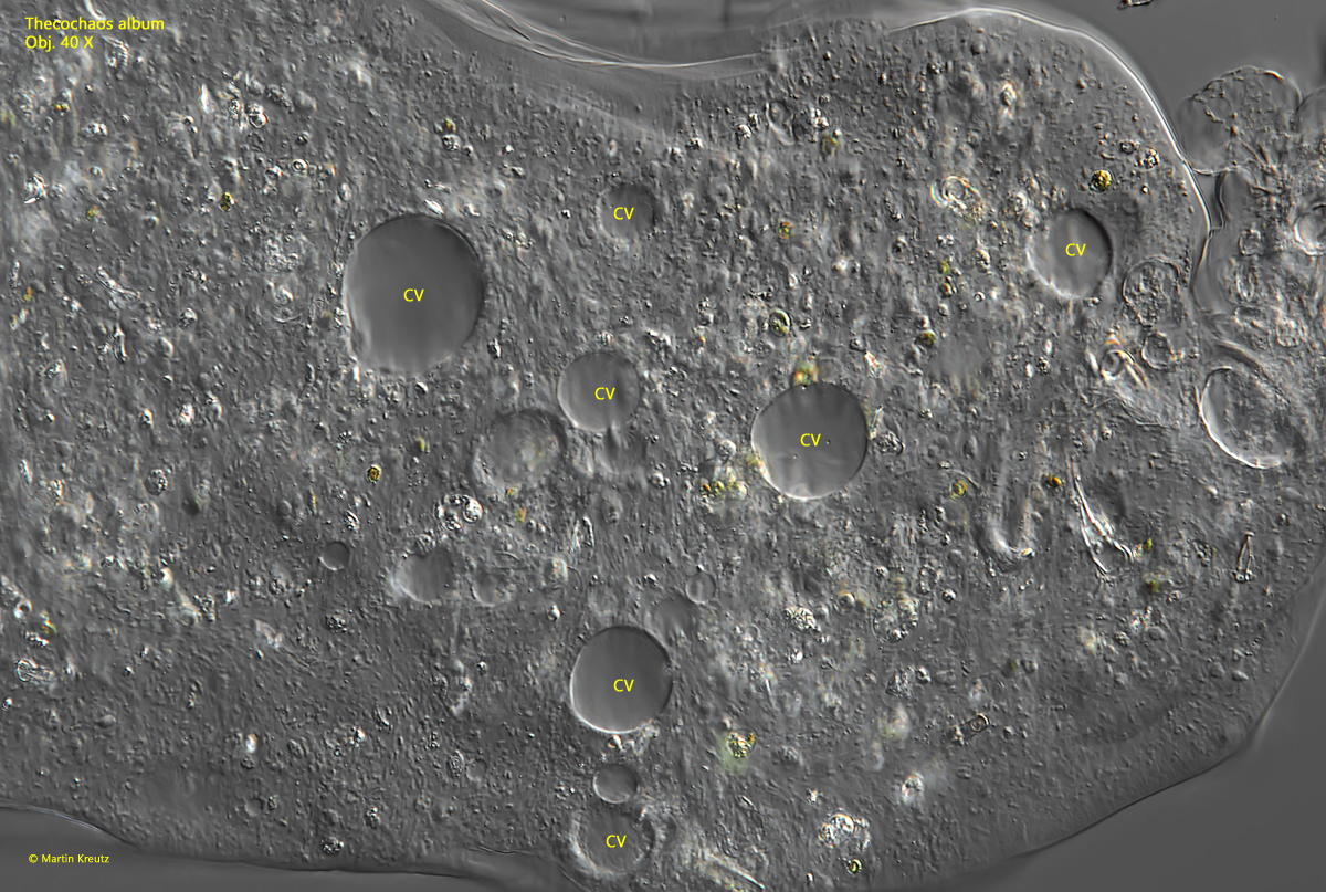
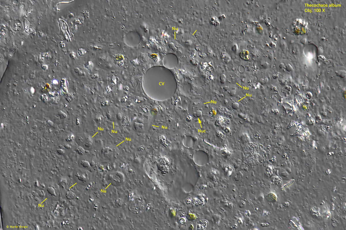
A thin rim of hyaline ectoplasm is formed at the front end during locomotion (s. fig. 4) and characteristic longitudinal wrinkles appear on the pellicle (s. fig. 1 b). These wrinkles constantly change in number and length, but are always present. I was able to recognize about 20 contractile vacuoles distributed in the cytoplasm (s. fig. 2 a-b). It is difficult to determine their exact number because of the constant change in their position and pulsating activity. Penard mentions a “considerable number” of vacuoles, although it remains unclear whether he meant contractile vacuoles.
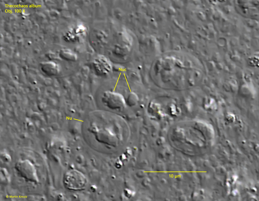
The main characteristics of Thecochaos are are the number and shape of the nuclei. Penard reported “several hundred” nuclei with an elliptical or round shape and a size of 7–12 µm (elliptical nuclei) or 6.5–7.5 µm (round nuclei). In my specimen, the nuclei were only recognizable in the squashed specimen, as their refractive index differed only slightly from that of the cytoplasm. I estimated their number at 100–300, which agrees well with Penard’s and Page’s data. In my specimen the nuclei were all broadly oval (s. figs. 5 and 6). All nuclei contained irregular nucleoli and had an average size of 7 x 9 µm. Often there appear to be two almost spherical nucleoli but also band-shaped and cloudy diffuse nucleoli. Penard described and drew several spherical nucleoli in each nucleus (s. drawing above). The shape of the nucleoli in Thecochaos album thus appears to be somewhat variable, but they are never central, spherical nucleoli, as in the similar species Thecochaos fibrillosum.
The cytoplasm was clear and highly refractive, similar to the genus Thecamoeba. I could not recognize any larger crystals in the cytoplasm. A few yellow-green food vacuoles were visible, which probably originated from phagocytized Oscillatoria chlorina. Penard found algae and diatoms in his specimens.