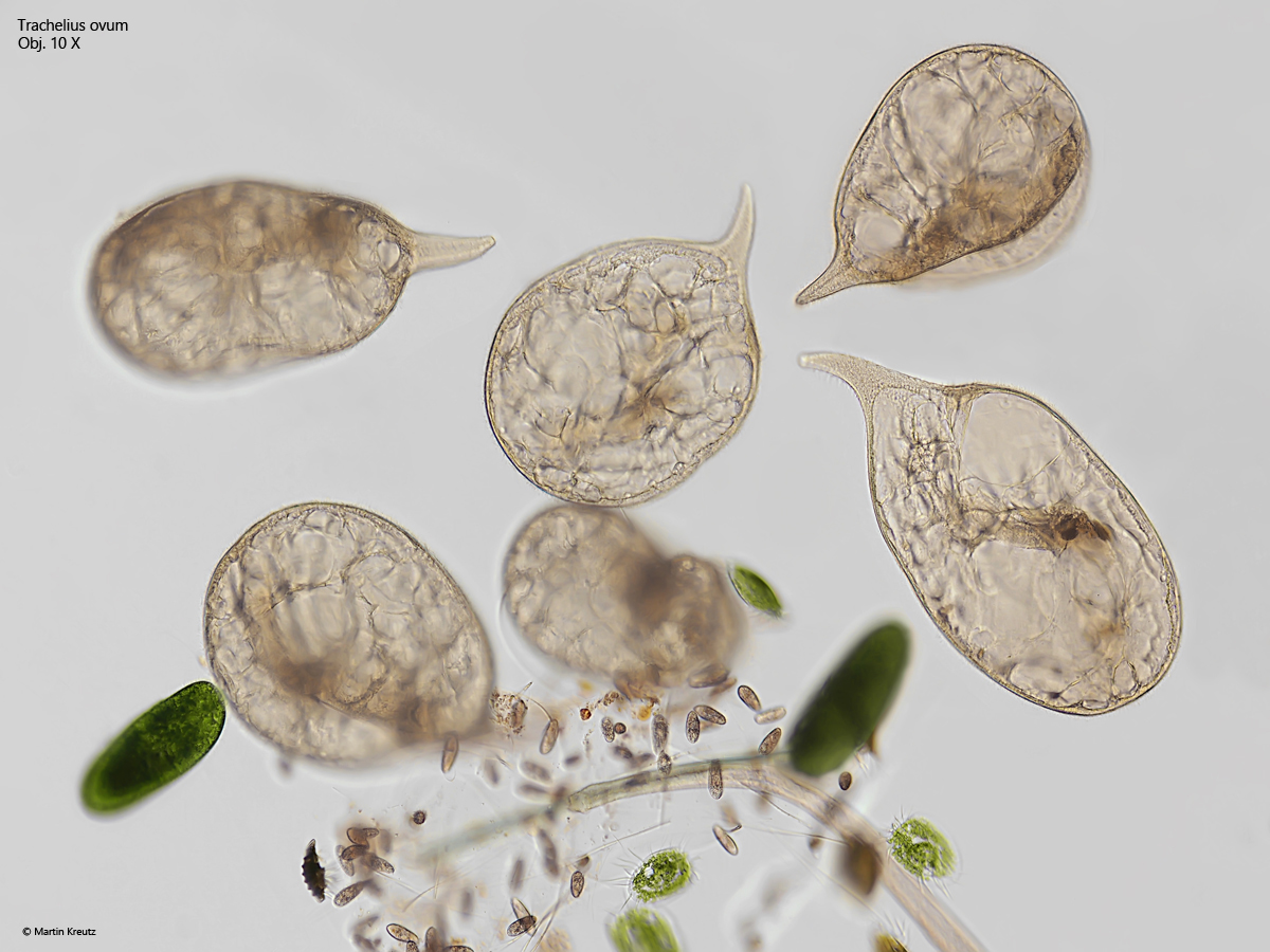
Trachelius ovum is a large and conspicuous ciliate that I find in many of my sampling sites. In old samples, it often multiplies when peritrich ciliates settle on the surface and on the vessel wall, as Trachelius ovum particularly likes to phagozytise these.
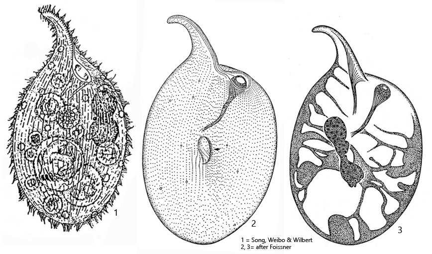
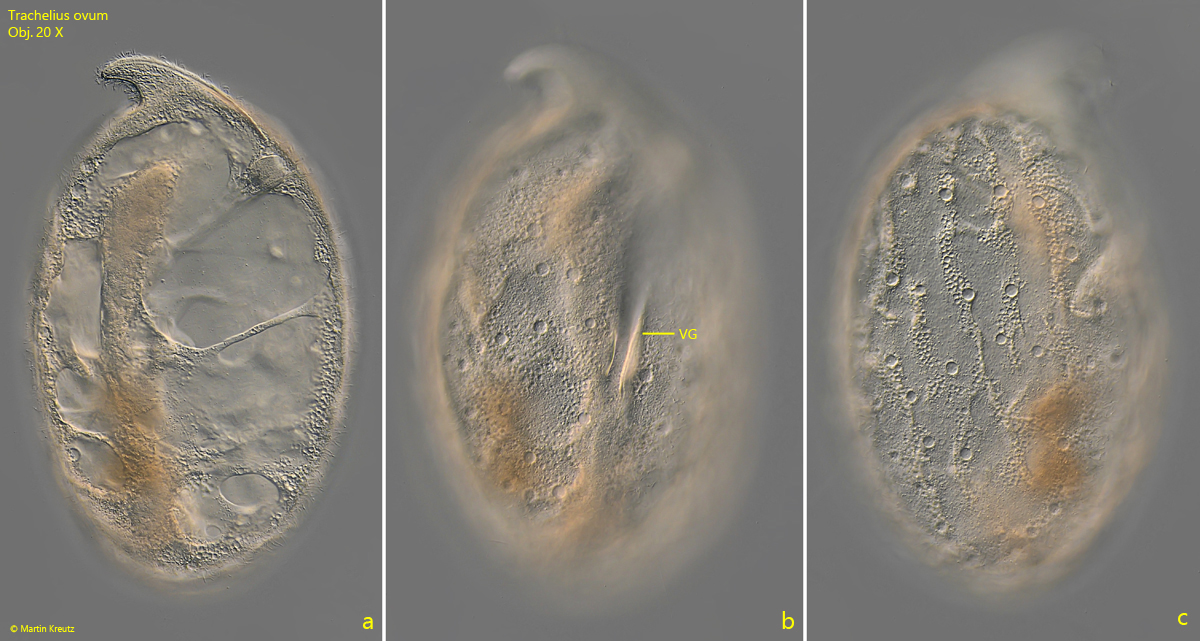
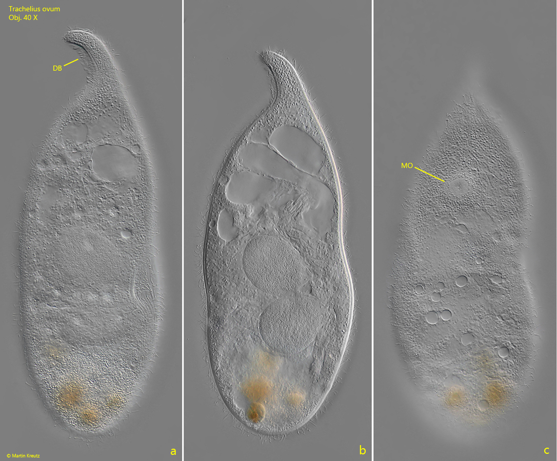
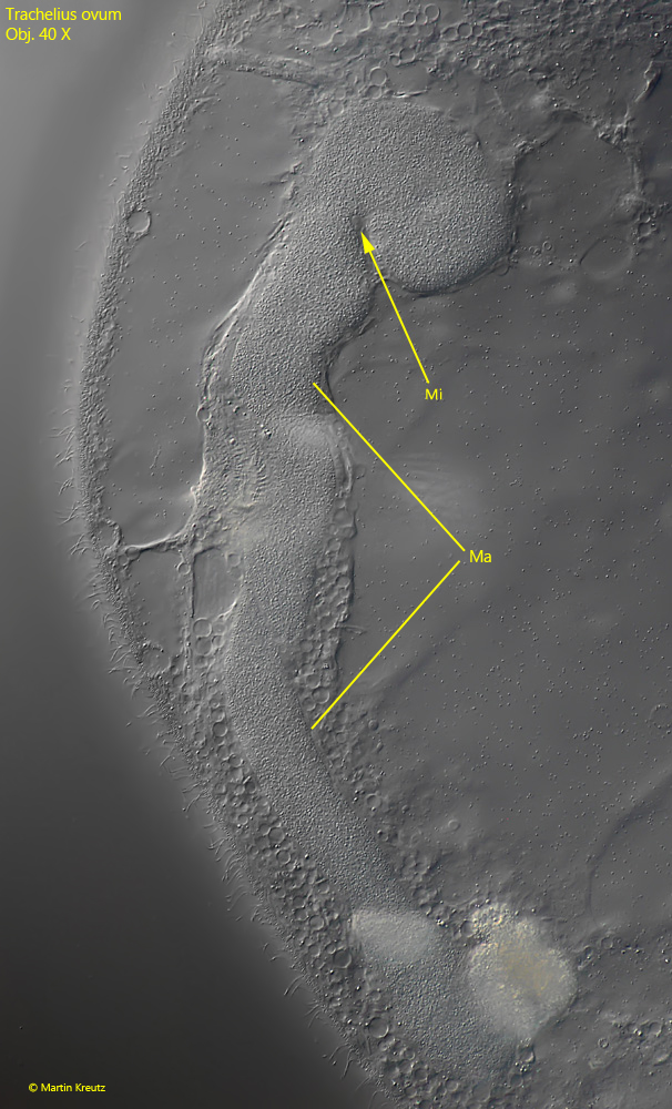
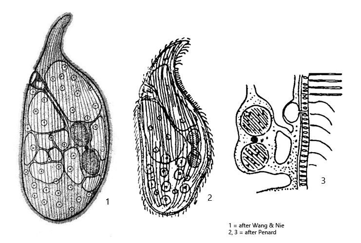
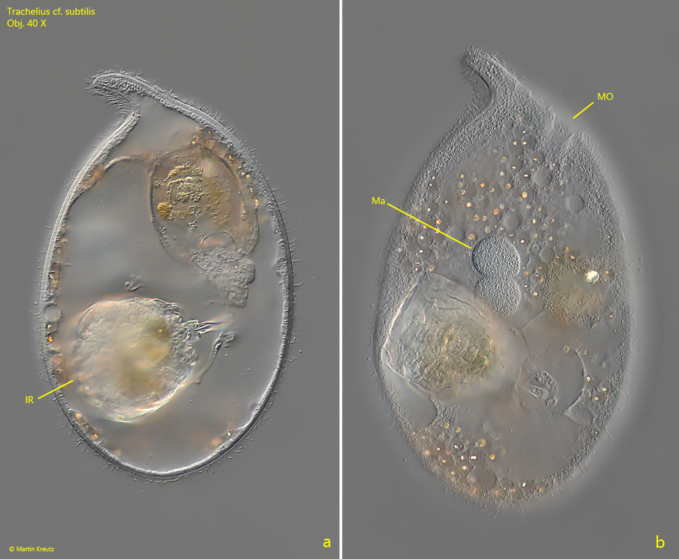
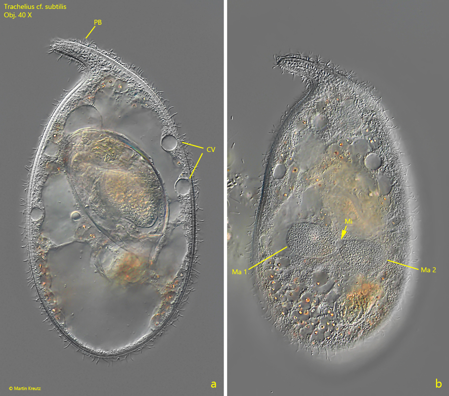
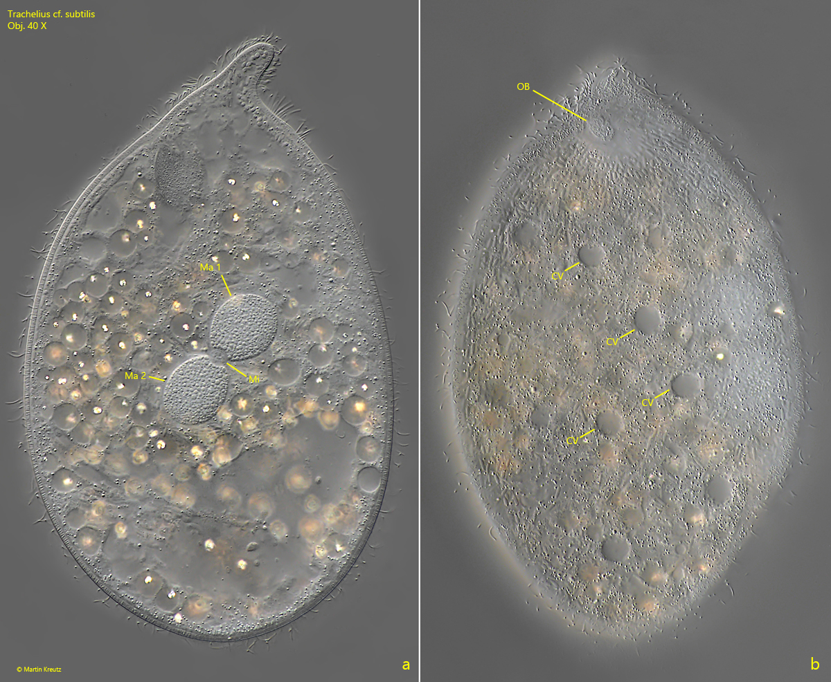
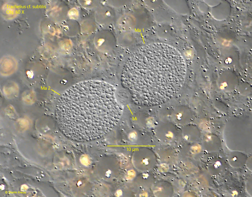
In brightfield illumination, the specimens mostly appear brown to brown-yellowish (s. fig. 1). The body shape is almost always plump and sack-shaped. In the specimens of my population, the proboscis was always quite short, about a quarter of the body length. The cytoplasm of Trachelius ovum is always strongly vacuolated, and in unsquashed specimens, the macronucleus is difficult to recognize because it is embedded in brownish cytoplasm. The shape of the macronucleus can vary greatly. Most often it is elongated and thread-like (s. fig. 6), but sometimes it is dumbbell-shaped or consists of several parts (s. fig. 3 b). The micronuclei are generally difficult to see and very small (s. fig. 6). There are supposed to be several.
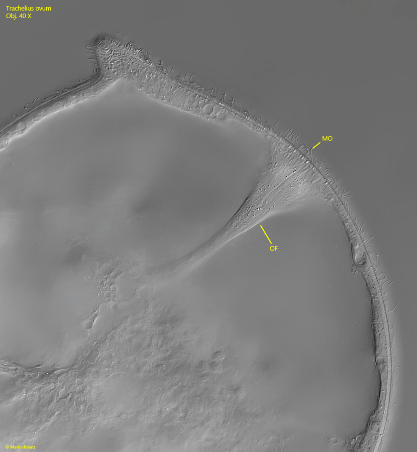
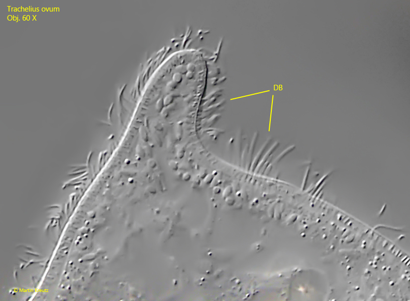
At the base of the proboscis lies the round or slightly elliptical mouth opening. It is surrounded by radially arranged rows of extrusomes, which extend upward in two parallel bands on the ventral side of the proboscis (s. fig. 7 a-b). Viewed laterally, the mouth opening is funnel-shaped, quickly narrowing and extending deep into the cytoplasm as a strand, similar to an esophagus (s. fig. 4).
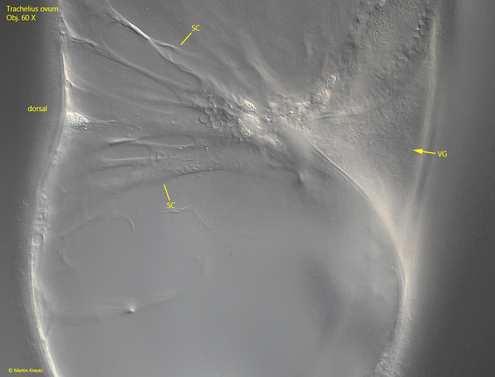
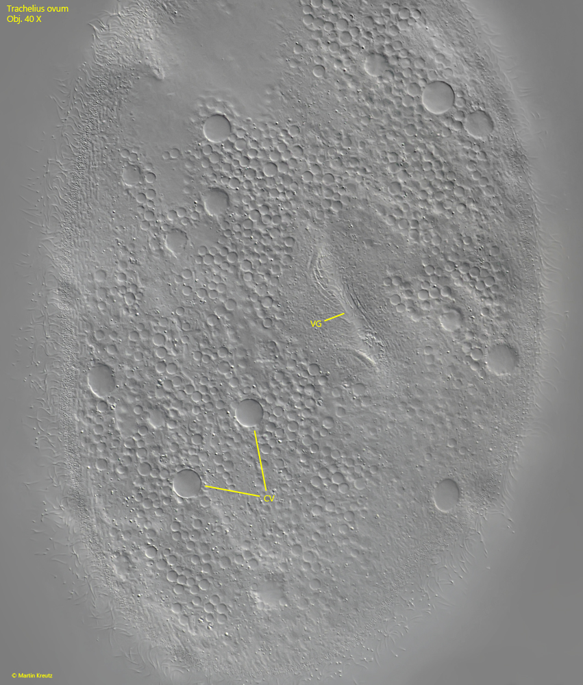
A unique feature of Trachelius ovum is a groove-shaped depression that is not entirely on the ventral side but is slightly shifted to the right side (s. fig. 2 b and 9). It is difficult to detect in freely swimming and rotating specimens. I was able to confirm the observation described by Kahl (1935) that the base of the groove is connected to the dorsal side by strands of cytoplasm, similar to muscle strands (s. fig. 5). The groove is formed and stabilized by the tension of the strands.
There are various theories about the function of this ventral groove. Hamburger (1903) is said to have observed specimens of Trachelius ovum clinging to the stalks of sessile peritrichs with the ventral groove and then detaching the zooids by rotating their bodies. However, this observation has not been confirmed by any other author since 1903. It is much more likely that the groove serves as a “fold for expansion.” When Trachelius ovum has phagocytosed large amounts of prey, the strands of cytoplasm expand and the groove serves as a volume reserve. This was also suspected by Kahl.
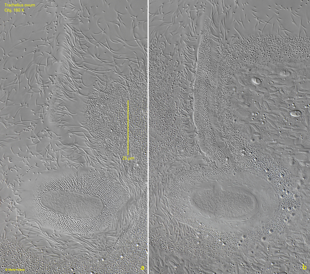
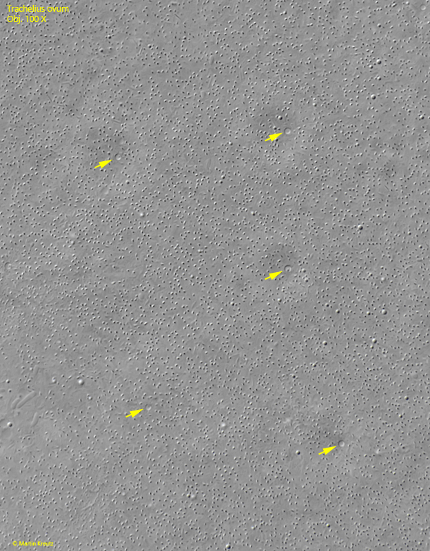
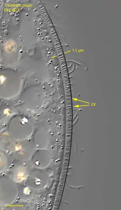
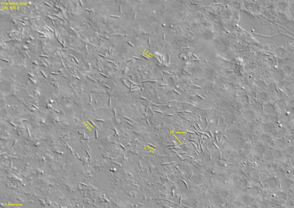
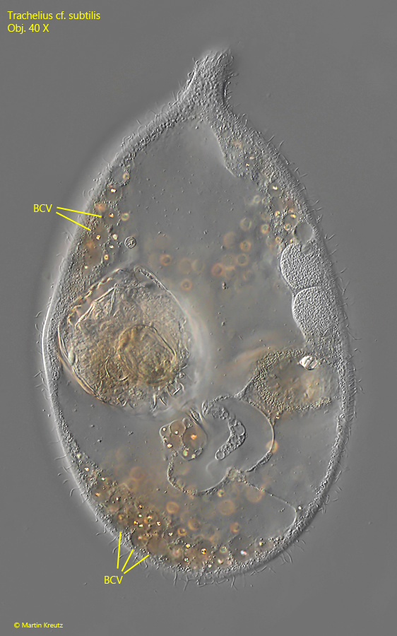
The body of Trachelius ovum is densely covered with very many contractile vacuoles (s. fig. 9). Each contractile vacuole has an excretory pore, which can also be clearly seen when focusing on the surface of the pellicle (s. fig. 10). The pellicle itself is quite thick and has a defined layer of short extrusomes about 1 µm long (s. fig. 11). They are different from the extrusomes located around the mouth opening and on the proboscis. According to my observations, these are also rod-shaped, but with slightly tapered ends and a length of 2-2.5 µm (s. fig. 12).