lorica brownish or orange, colorless in young specimens
lorica with long spine at posterior end and towards the collar
equatorial zone with short spines
cell with 8–10 disc-shaped chloroplasts without pyrenoid
one eyespot
flagellum 1–2 times of body length
pellicle with striation running in counterclockwise direction
Trachelomonas hystrix
Trachelomonas hystrix is a very constant species in the Simmelried. I have not yet been able to find Trachelomonas hystrix in my other sampling sites.
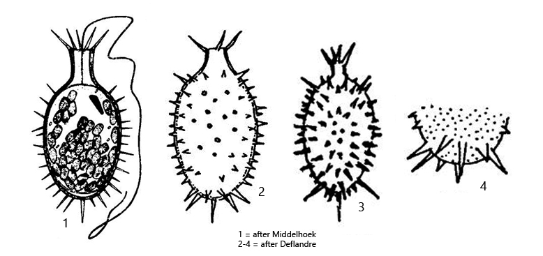
Trachelomonas hystrix is very conspicuous due to its spiny lorica. These can be divided into four groups:
– the spines on the margin of the collar – the elongated spines in the shoulder area below the collar – the short spines in the equatorial zone – the long spines at the posterior end
The lorica itself is mostly brownish or orange in color due to the inclusion of iron or manganese salts. Only young specimens are initially almost colorless. Only in such specimens can the protoplast with the disc-shaped chloroplasts and the striation of the pellicle be examined (s. fig. 4 a-b). In my population the specimens had 20–30 chloroplasts and not only 8–10 as described by earlier authors. I could not observe any pyrenoids. The flagellum of my specimens was also always shorter, at most as long as the body.
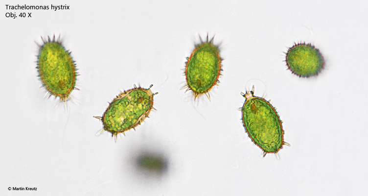
Fig. 1:Trachelomonas hystrix. L = 39–41 µm (from margin of collar to rounded base of lorica). Some freely swimming specimen. Obj. 40 X.
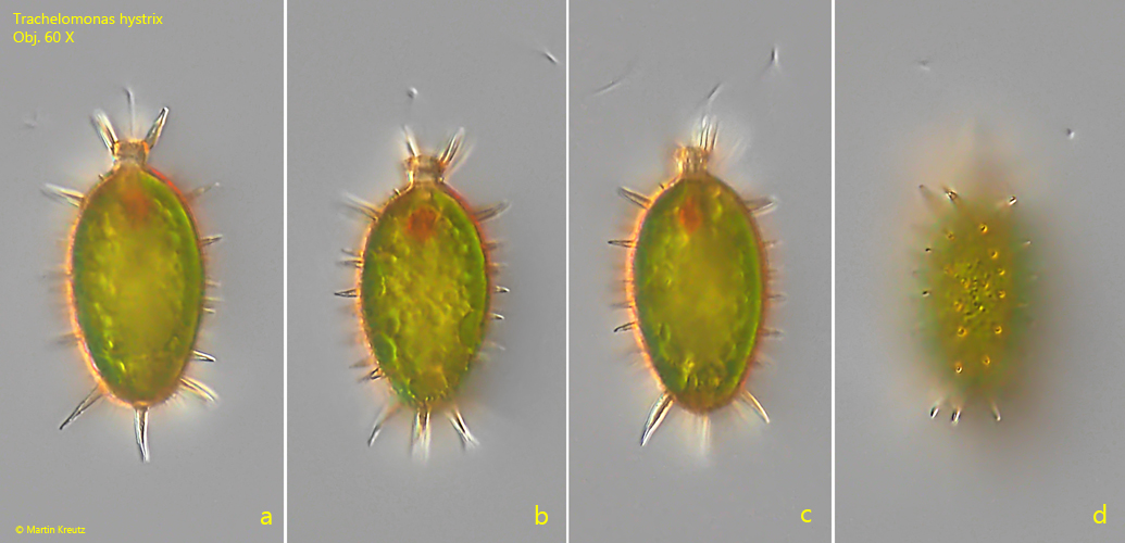
Fig. 2 a-d:Trachelomonas hystrix. L = 42 µm (from margin of collar to rounded base of lorica). Different focal planes of a freely swimming specimen. Obj. 60 X.
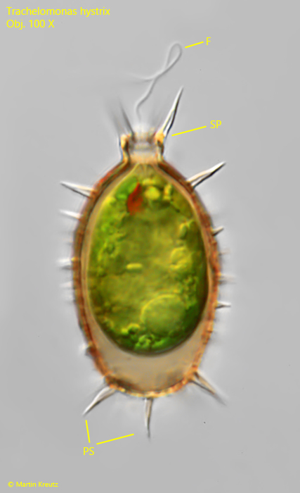
Fig. 3:Trachelomonas hystrix. L = 41 µm (from margin of collar to rounded base of lorica). A freely swimming specimen in detail. Note the spiny collar and the elongated spines at the posterior end (PS) while the spine in the equatorial zone are shorter. F = flagellum. Obj. 100 X.
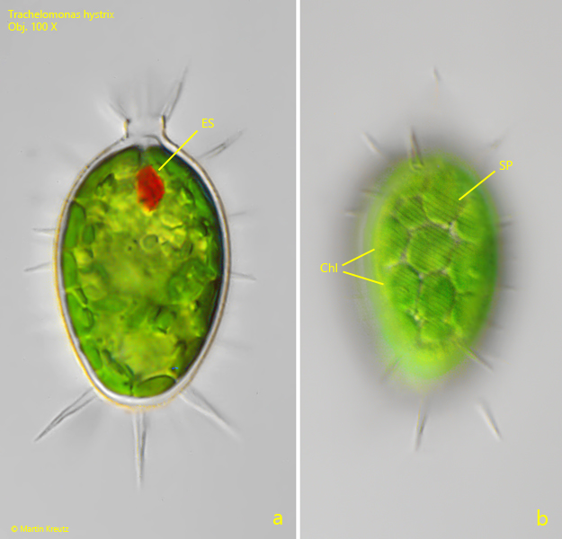
Fig. 4 a-b:Trachelomonas hystrix. L = 38 µm (from margin of collar to rounded base of lorica). Two focal planes of a young, transparent specimen. Note the disc-shaped chloroplasts (Chl) and the striation of the pellicle (SP) running in counterclockwise direction. ES = eyespot. Obj. 100 X.
Fig. 5 a-b:Trachelomonas hystrix. L = 47 µm (from margin of collar to rounded base of lorica). Two focal planes of a specimen in brightfield illumination. Obj. 100 X.
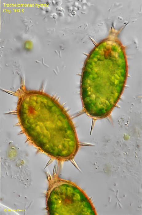
Fig. 6:Trachelomonas hystrix. L = 36–42 µm (from margin of collar to rounded base of lorica). Three older specimens with orange-brownish colored loricae and strongly developed spines. Obj. 100 X.