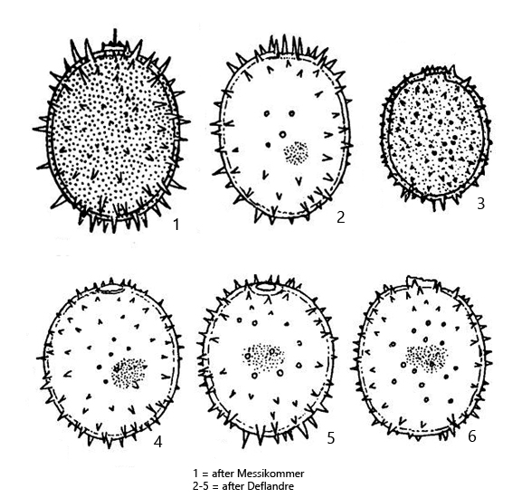
lorica broadly ellipsoidal to ovoid, rounded apices
apical pore without collar, sometimes with a thickened bulge
lorica punctuated and completely covered with conical spines
spines somtimes longer around apices
length 38–55 µm, width 30–39 µm
lorica brownish or orange
eyespot large
chloroplasts disc-shaped
flagellum about body length
Trachelomonas superba
Trachelomonas superba is one of the most common representatives of the genus in the Simmelried. I have not yet been able to find this species in my other sampling sites.
Trachelomonas superba can be easily recognized by the conical spines, which are evenly distributed with larger distances on the lorica. These loosely spaced, conical spines distinguish Trachelomonas suberba from the similar species Trachelomonas hispida, which has much shorter spines that are much closer together. In addition, Trachelomonas hispida rarely grows longer than 30 µm.
In my population, the specimens of Trachelomonas superba reached a length of over 40 µm without exception. In contrast to the descriptions of earlier authors, the flagellum of my specimens was significantly longer than the body, up to twice the body length. Many specimens had a distinct mucus layer (s. fig. 4 a-b), which is not mentioned in the literature.
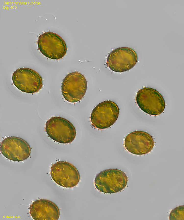
Fig. 1:Trachelomonas suberba. An accumulation of freely swimming specimens. Obj. 40 X.
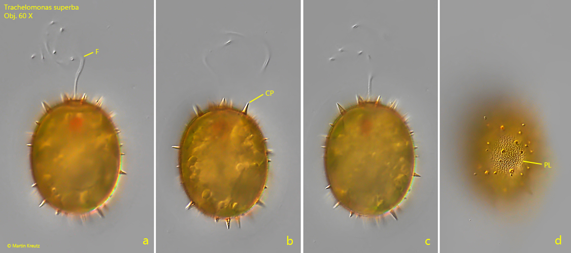
Fig. 2 a-d:Trachelomonas suberba. L = 46 µm (without spines). Different focal planes of a freely swimming specimens. CP = conical spines, F = flagellum, PL = punctuated lorica. Obj. 60 X.
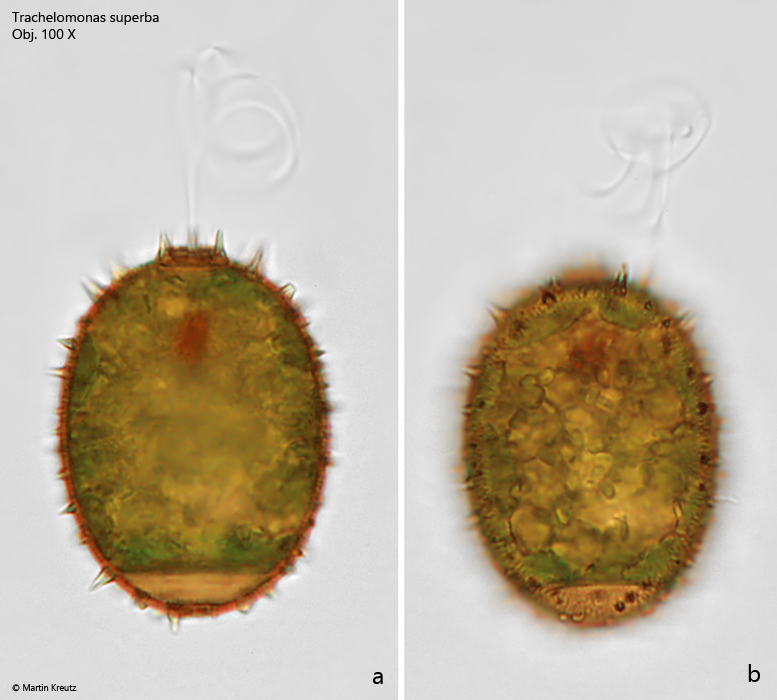
Fig. 3 a-b:Trachelomonas suberba. L = 48 µm (without spines). Two focal planes of a freely swimming specimen in brightfield illumination. Obj. 100 X.
Fig. 4 a-b:Trachelomonas suberba. L = 28 µm (without spines). Two focal planes of a freely swimming specimen with a clearly visible mucus layer (ML). Obj. 100 X.
Fig. 5 a-b:Trachelomonas suberba. L = 28 µm (without spines). The same specimen as shown in fig. 4 a-b in brightfield illumination. Obj. 100 X.
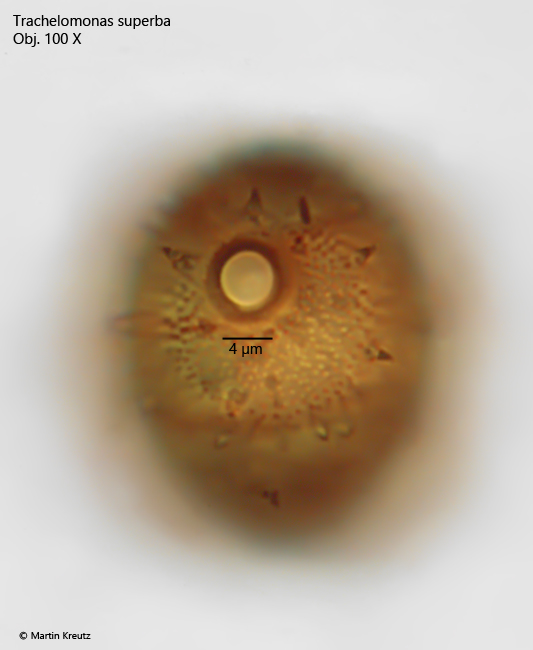
Fig. 6:Trachelomonas suberba. Apikal view of the circular pore with a diameter of 4 µm. The pore is surrounded by a thickened bulge. Obj. 100 X.