posterior end with a distinct cytoplasmic spine, directed ventrally
Trochilia minuta
Trochilia minuta is a very common ciliate, which I find in almost all my localities. However, it can be easily overlook in the samples because the specimens are usually much smaller than 30 µm.
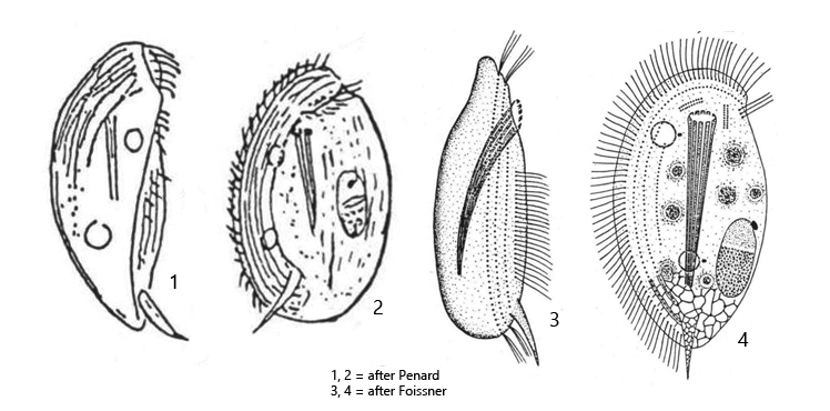
Trochilia minuta has a complex body structure as an adaptation to its lifestyle. The ciliate is often found running over detritus flakes or filaments of cyanobactria and green algae, similar to a turtle, in order to feed on bacteria adhering to them. Locomotion is by means of a ventral ciliated band, which only covers the right half of the body. The mouth opening is surrounded by cyoplasmic lips and the pharynx is a tube of trichites (= oral basket) that extends to the unciliated dorsal side (s. fig. 1).
Very characteristic is a cytoplasmic spine at the posterior end, which usually points ventrally (s. figs. 1 , 3 a and 6b). I had the impression that it serves as a tactile organelle, as the spine is constantly in contact with the substrate when the ciliate runs over surfaces. I was also able to observe that the cytoplasmic lips surrounding the mouth opening can apparently be stretched far forward and then, similar to a proboscis, seem to scan the surface of the substrate (s. fig. 2 b). To my knowledge, this behavior has not yet been described. In one case I was also able to observe phagocytosis (s. below).
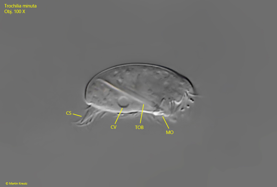
Fig. 1:Trochilia minuta. L = 20 µm. Lateral view from right of a slightly squashed specimen. Note the distinc cytoplasmic spne (CS) at the posterior end. CV = one of the two contractile vacuoles, MO = mouth opening, TOB = trichites of oral basket. Obj. 100 X.
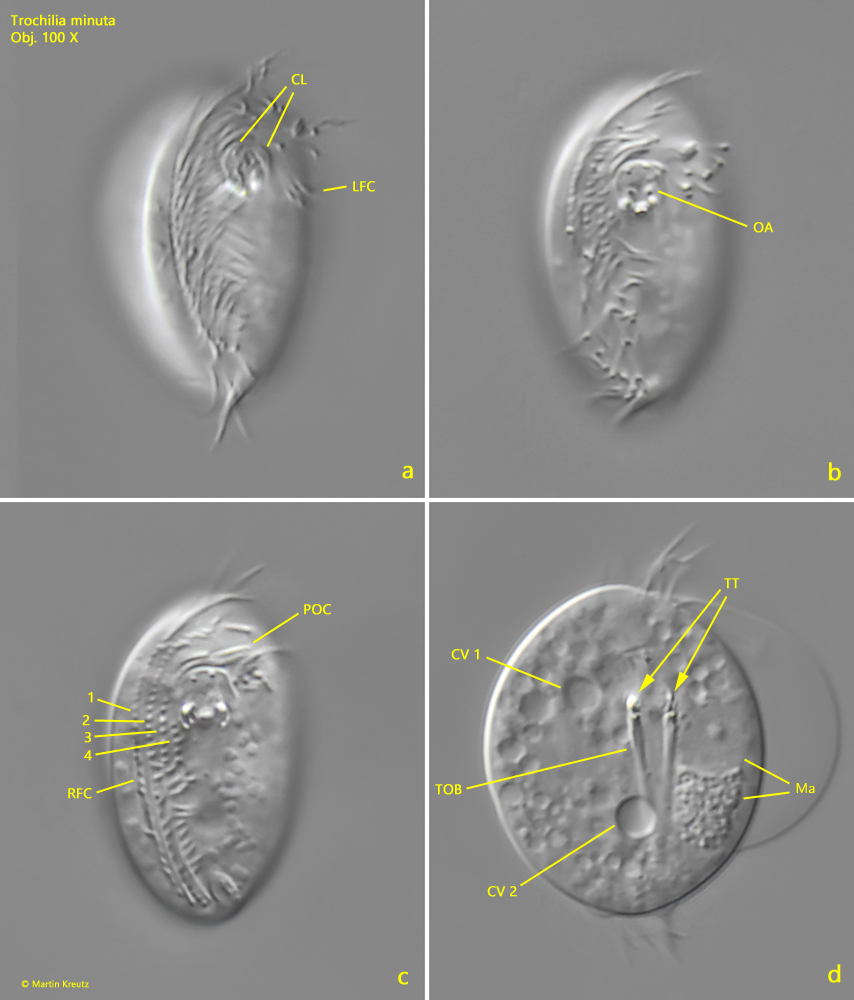
Fig. 2 a-d:Trochilia minuta. L = 23 µm. Different focal planes of a slightly squashed (a-c) and strongly squashed (d) specimen from ventral. Only the right side is ciliated with 4 rows of cilia (1-4) forming the right field of cilia (RFC). At the left side, the very small left field of cilia is visible (LFC). Above the mouth opening the two rows of pre-oral cilia (POC) are visible. The aral apparatus (OA) is surrounded by cytoplasmic lips (CL). The trichites of the oral basked have small teeth at the anterior ends (TT). CV = contractile vacuoles, Ma = macronucleus. Obj. 100 X.
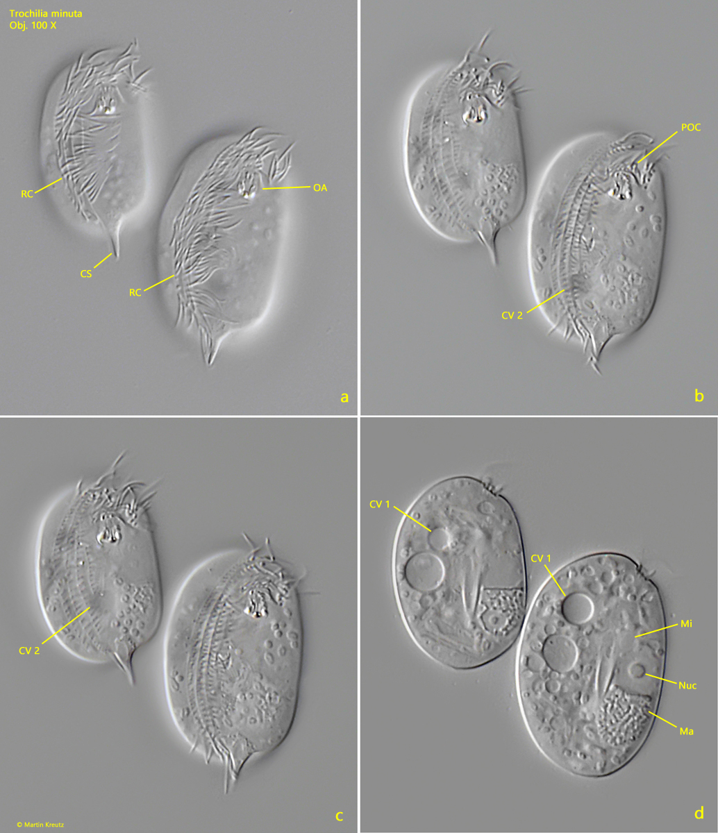
Fig. 3 a-d:Trochilia minuta. L = 25–28 µm. Two slightly squashed specimens from ventral. Note the spherical nucleolus (Nuc) in the homogeneous half of the macronucleus (Ma). CV 1, CV 2 = contractile vacuoles, CS = cytoplasmic spine, Mi = micronucleus, OA = oral apparatus, POC = pre-oral rows of cilia, RC = right field of cilia. Obj. 100 X.
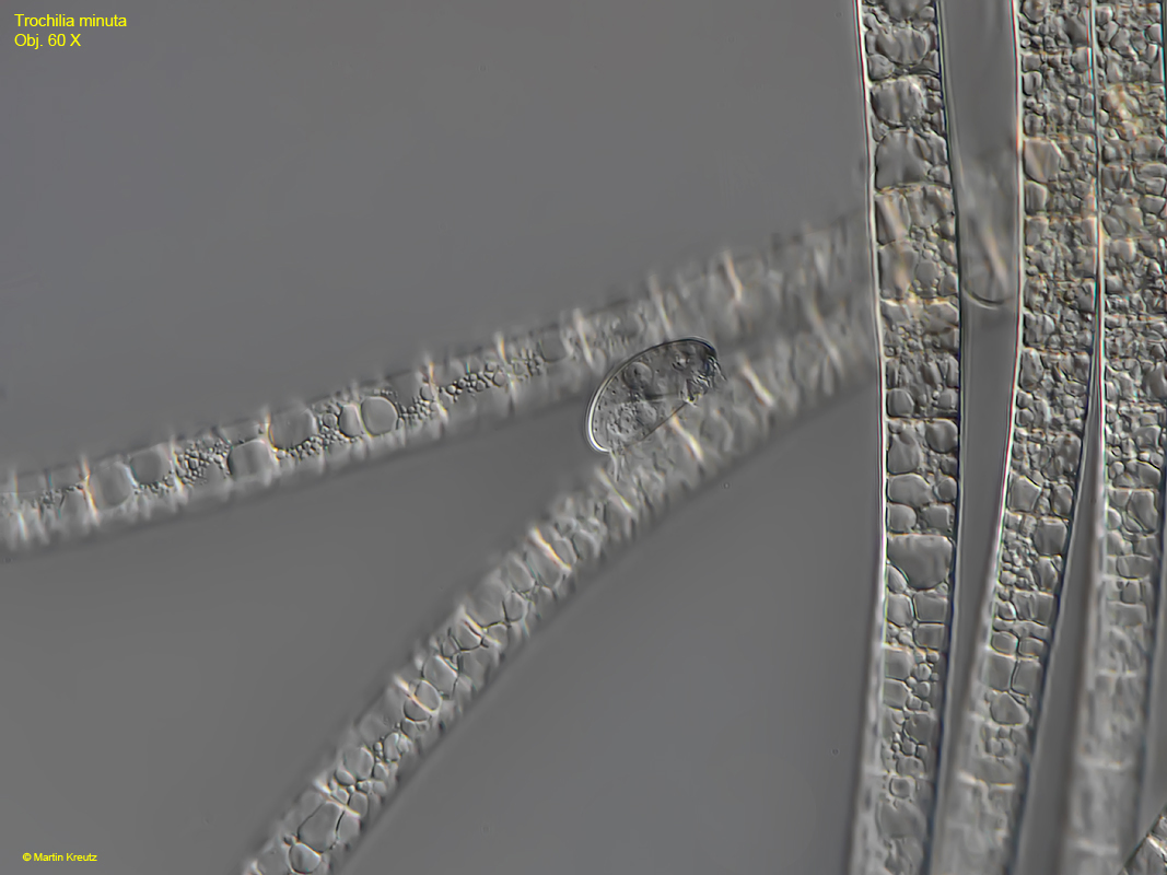
Fig. 4:Trochilia minuta. L = 24 µm. A specimen is crawling over the filaments of the cyanobacterium Tychonema spec. Obj. 60 X.
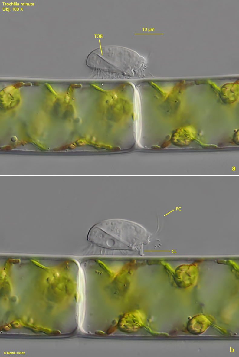
Fig. 5 a-b:Trochilia minuta. L = 22 µm. A specimen is crawling over the filaments of Spirogyraspec. Note the stretched out cytoplasmic lips (CL) similar to a trunk. TOB = trichites of oral basket. Obj. 100 X.
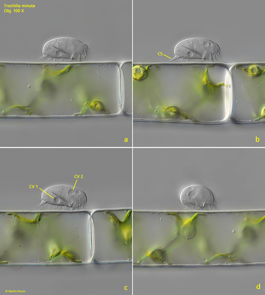
Fig. 6 a-d:Trochilia minuta. L = 22 µm. A second specimen specimen crawling over the filaments of Spirogyraspec. from right (a–c) and from anterior (d). Note the two contractile vacuoles (CV 1, CV 2). CS = cytoplasmic spine. Obj. 100 X.
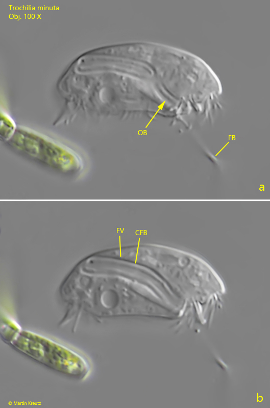
In one case I had the opportunity to observe Trochilia minuta feeding (see fig. 7 a-b). The specimen was phagocytizing a filamentuous bacterium. Before doing so, it obviously felt this filamentuous bacterium to find an end. Unfortunately, I could not see whether the cytoplasmic lips were used in the process. When the specimen found the end of the filemant, the phagocytosis began immediately, which lasted only a few seconds (about 10–15 seconds). It was clearly visible how the filamentuous bacterium passed the oral basket (s. fig. 7 a) and wound up in an elongated food vacuole (s. fig. 7 b).
Fig. 7 a-b:Trochilia minuta. L = 27 µm. A specimen is feeding on a filamentuous bacterium (FB). The bacterium passed the oral basked (OB) and was wound up in an elongated food vacuole (FV). Obj. 100 X.