body elongate ellipsoidal, strongly spirally furrowed by 7–8 cortical ridges, shape very variable
anterior end obliquely truncate, posterior end with distinct spine
length 70–180 µm
adoral zone short (11–16 membranelles), right anteriorly
perizonal stripe longer than adoral zone
7–8 rows of cilia running in the furrows of pellicle
somatic cilia arranged in pairs
macronucleus elongate ellipsoidal with adjacent, spherical micronucleus
contractile vacuole at base of spine
Tropidoatractus acuminatus
I find Tropidoatractus acuminatus regularly in the Purren pond and the Simmelried. The occurrence of this ciliate seems to be quite different. For example, Foissner describe the occurrence as “very rare”, while Kahl describes it as “common, never frequent”.
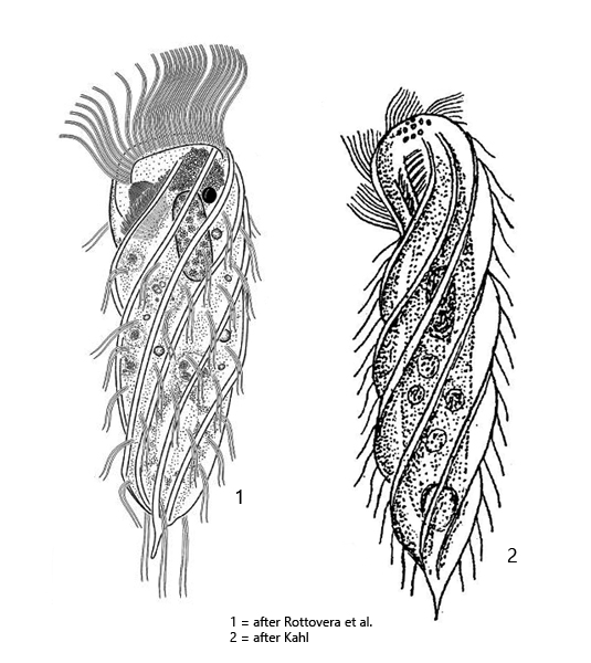
Tropidoatractus acuminatus is clearly and easily identifiable by its 7–8 distinct ribs, which running clockwise over the body (s. figs. 1 a and 2 e). The posterior end is elongated to a pointed spine (s. fig. 2 d). The specimens are usually longer than 100 µm. However, Tropidoatractus acuminatus is known to be very rich in form. Thus, also strongly broadened, stocky forms can be found and those with a very long terminal spine. In addition, dwarf forms of Tropidoatractus acuminatus can be found (see below).
The anterior end is flattened and curved ventrally. On the anterior margin of the apikal dome runs the perizonal stripe, with densely standing, long cilia (s. fig. 2 c). The somatic cilia are arranged in pairs between the ribs (s. fig. 5). In my population the macronucleus was usually quite long and ellipsoidal (s. fig. 4). It was mostly found in the anterior third of the body with an attached, spherical micronucleus (s. fig. 4). I could not detect any symbiotic bacteria in the cytoplasm and none have been described so far. The adoral zone is located on the right side at the anterior end. It is partially covered by the ribs. From the right side, however, it can be seen well (s. fig. 2 a and 2 b).
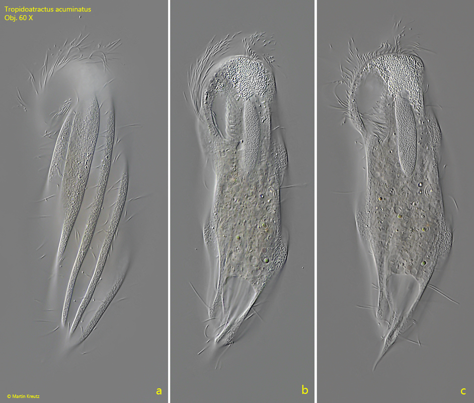
Fig. 1 a-c:Tropidoatractus acuminatus. L = 126 µm. Three focal planes of a freely swimming specimen from ventral. Obj. 60 X.
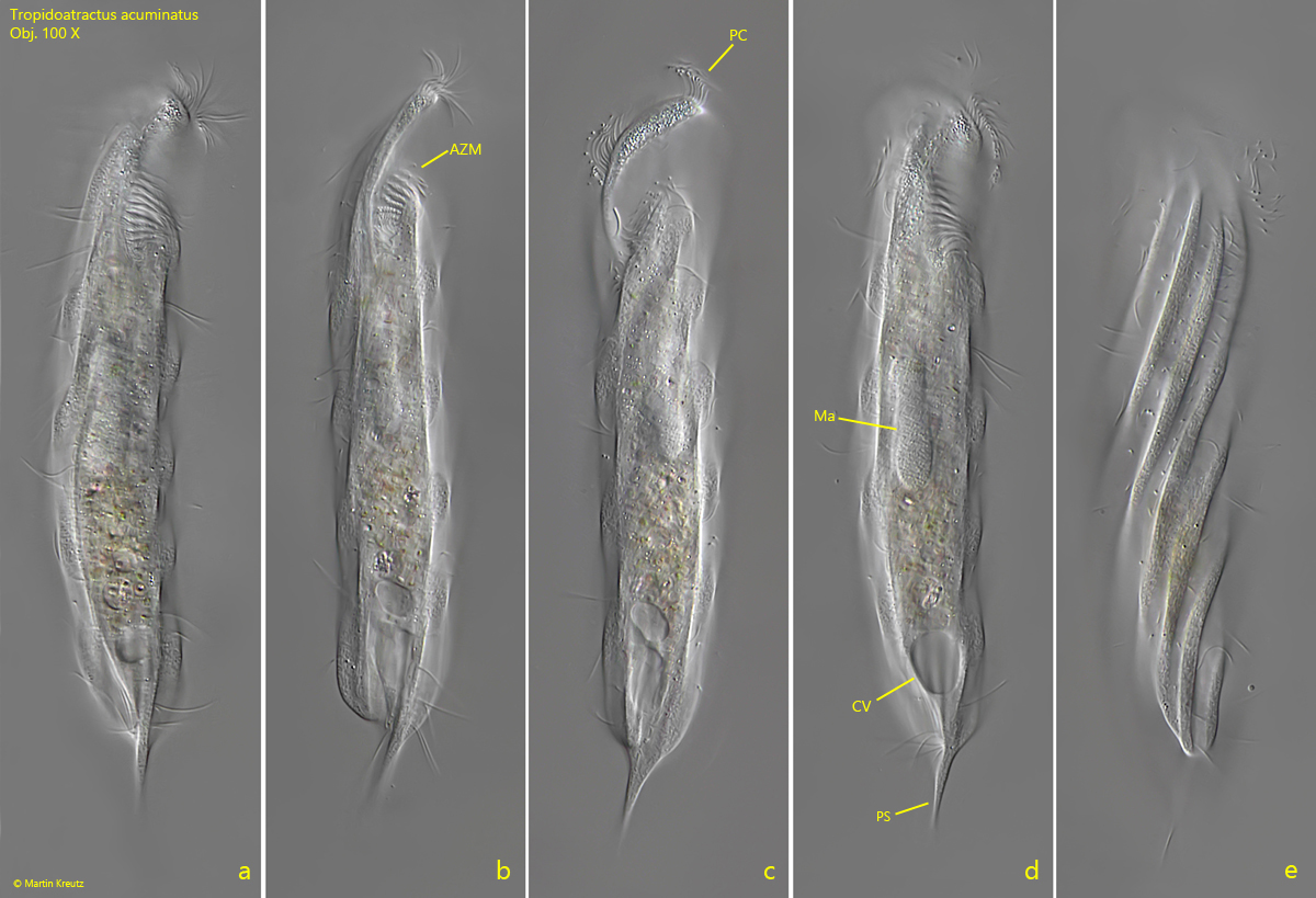
Fig. 2 a-e:Tropidoatractus acuminatus. L = 142 µm. A second freely swimming specimen from right (a, b, d) and ventral (c, e). AZM = adoral zone of membranelles, CV = contractile vacuole, Ma = macronucleus, PC = perizonal cilia, PS = posterior spine. Obj. 100 X.
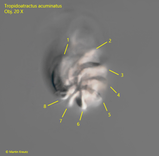
Fig. 3:Tropidoatractus acuminatus. Apical view from the posterior end. Note the 8 ribs of this specimen (1–8) running clockwise to the anterior end. Obj. 20 X.
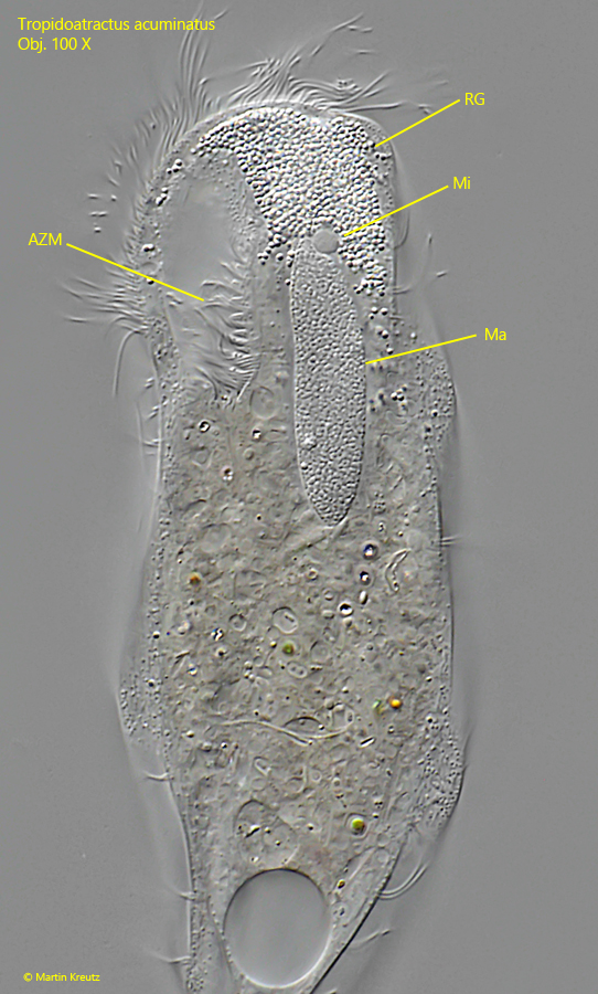
Fig. 4:Tropidoatractus acuminatus. The macronucleus (Ma) with the adjacent micronucleus (Mi) in a strongly squashed specimen. AZM = adoral zone of membranelles, RG = accumulation of refractive granules in the apical dome. Obj. 100 X.
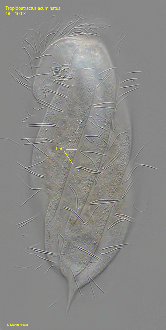
Fig. 5:Tropidoatractus acuminatus. Focal plane on the ventral somatic ciliation of a strongly squashed specimen. The cilia are widely spaced and arranged in pairs (PoC). Obj. 100 X.
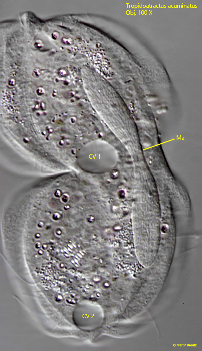
Fig. 6:Tropidoatractus acuminatus. A specimen during cell division. CV 1, CV 2 = contractile vacuoles, Ma = macronucleus in division. Obj. 100 X.
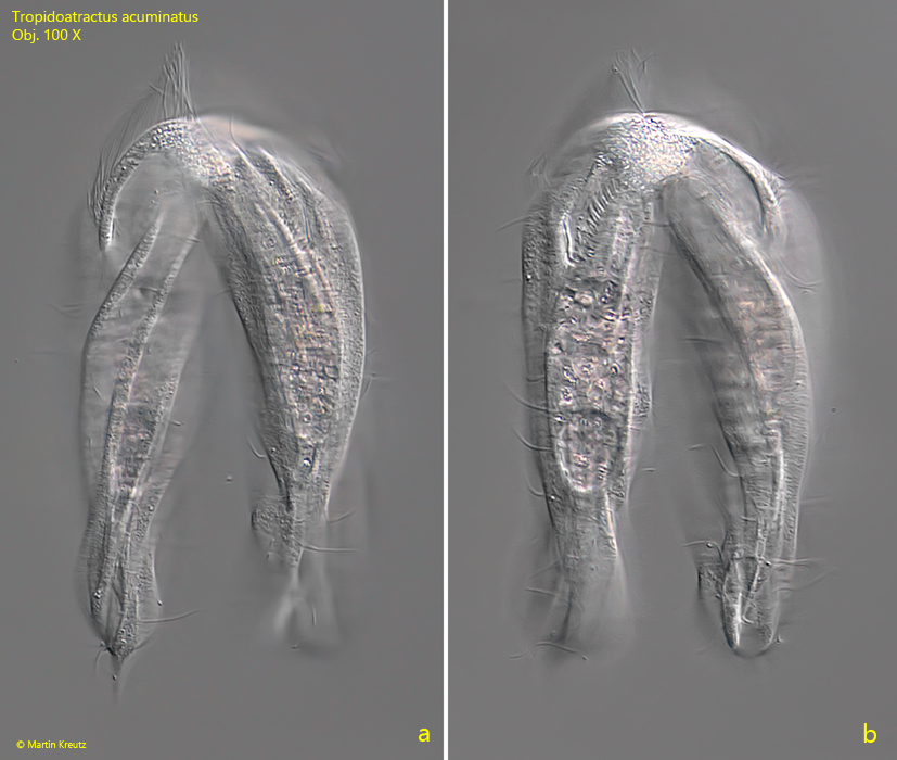
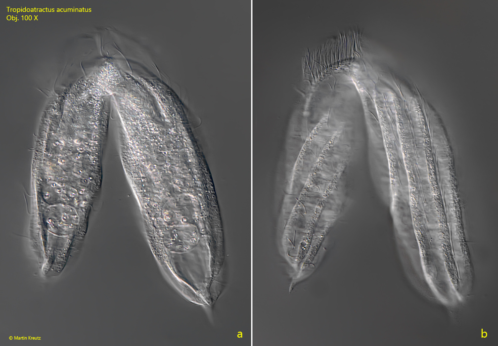
Tropidoatractus acuminatus can be observed very often during conjugation. Thereby the specimens fuse with the apikal domes and not via the mouth openings. In the area were the specimens are connected an accumulation of refractive granules is visible. It seemed to me that the pellicle of the two specimens also merged and formed a continuous apical arch on which a continuous perizonal stripe ran (s. fig. 8b).
Fig. 7 a-b:Tropidoatractus acuminatus. Two focal planes of a pair in conjugation. Obj. 100 X.
Fig. 8 a-b:Tropidoatractus acuminatus. A second pair in conjugation. Obj. 100 X.
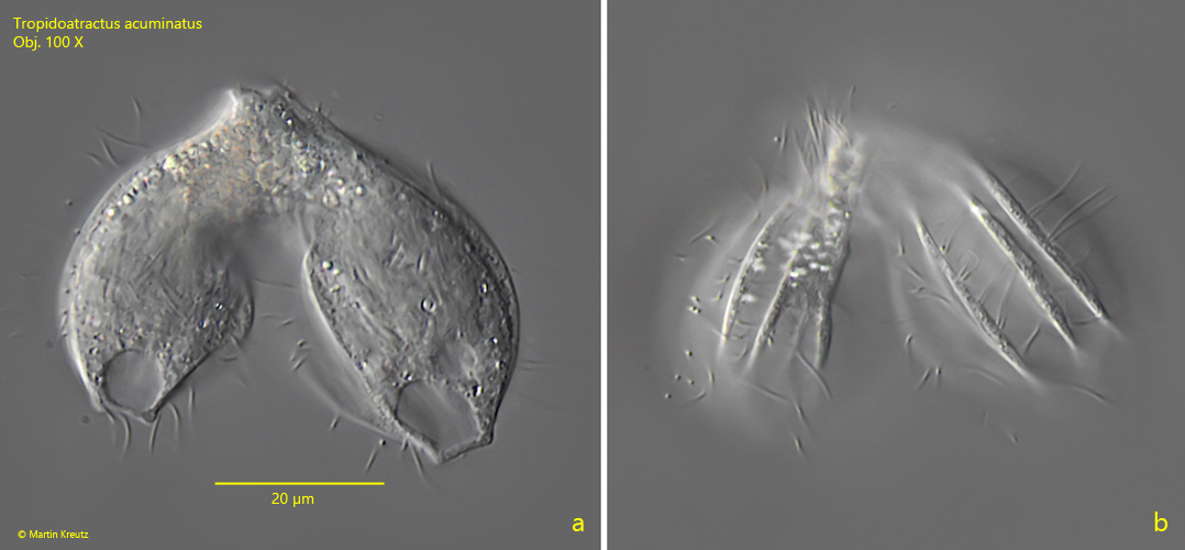
Already by Penard and Kahl dwarf forms of Tropidoatractus acuminatus were described, in which the terminal spine is reduced. I could also find these forms frequently. Some had a clearly truncated posterior end. The furrows of the pellicle did not run spirally in these forms, but rather straight over the whole body. The length of these forms was oftenl below 50 µm and I found them frequently in conjugation (s. fig. 9 a-b).
Fig. 9 a-b:Tropidoatractus acuminatus. A pair of dwarf forms with lengths of 38 µm and 44 µm in conjugation. Obj. 100 X.