body slightly S-shaped with a caudal spine fourth of body length
caudal spine delimited from right or left edge of body
length 50–115 µm
macronucleus oval, filled with symbiotic bacteria
one spherical micronucleus adjacent to the macronucleus
contractile vacuole subterminal, at the base of the caudal spine
accumulation of refractive granules in apical dome (often brownish)
adoral zone running obliquely to anterior fourth, not reaching equator
perizonal cilia row in parallel to adoral zone
caudal spine ciliated, but no elongated caudal cilia
Tropidoatractus spinosus
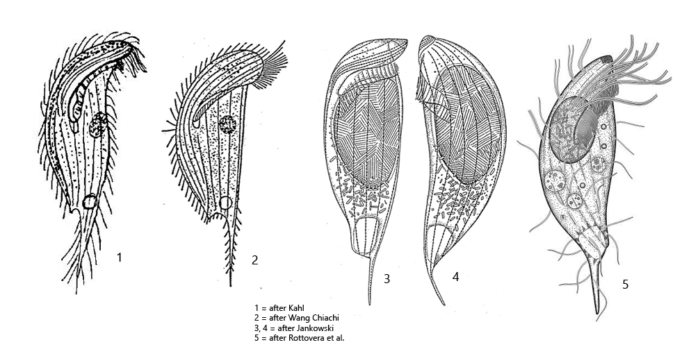
Tropidoatractus spinosus was first described as Metopus spinosus by Kahl in 1927. In 2018, Rottovera et al. redescribed the species and transferred it to the genus Tropidoatractus based on morphological characters.
In my localities Simmelried and Bündtlisried I find Tropidoatractus spinosus rarely, but regularly. The species is easily recognized by the slightly twisted, S-shaped body and the typical caudal spine, which is shifted to one side of the body. In my population I could find some differences to the descriptions of Tropidoatractus spinosus I have. On all drawings (apart from Rotterova et al., s drawings above) of Tropidoatractus spinosus the caudal spine is drawn on the left side of the body. In my specimens, however, it was localized exclusively on the right body margin (s. figs. 1 a-c and 2 a-c). My specimens were up to 100 µm long (s. fig. 2 a-c) and thus much longer than Kahl indicates (50–80 µm) but in the range indicated by Rottovera et al. (60–115 µm). Very striking in all specimens I examined was that the macronucleus was completely filled with at least two species of bacteria (s. fig. 3 a-b), whereas the cytoplasm was free from bacteria. According to Rotterova et al. these bacteria are presumably parasitic comparable to Holospora in the micronucleus of Paramecium. However, in all specimens of Tropidoatractus spinosus I observed, the macronucleus was infected with these bacteria. This makes a parasitic nature of the bacteria very unlikely, because an infection rate of 100% does not occur even with Holospora. The two types of bacteria are not homogeneously distributed in the macronucleus. In the margin of the macronucleus thin rods with a length of about 4 µm are localized (s. fig. 3a), while in the center there are mainly thicker rods with a length of about 5 µm (s. fig. 3b).
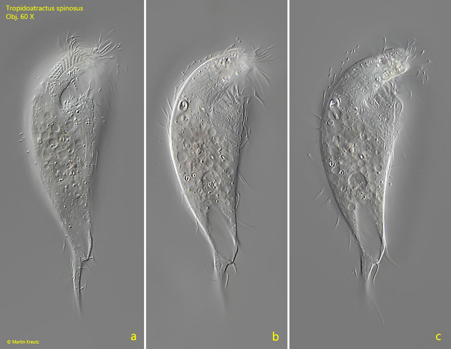
Fig. 1 a-c: Tropidoatractus spinosus. L = 94 µm. Ventral view of a freely swimming specimen. Note that the caudal spine is oriented on the right side of the body, which contrasts with the drawings by Kahl and other authors (s. drawings above). Obj. 60 X.
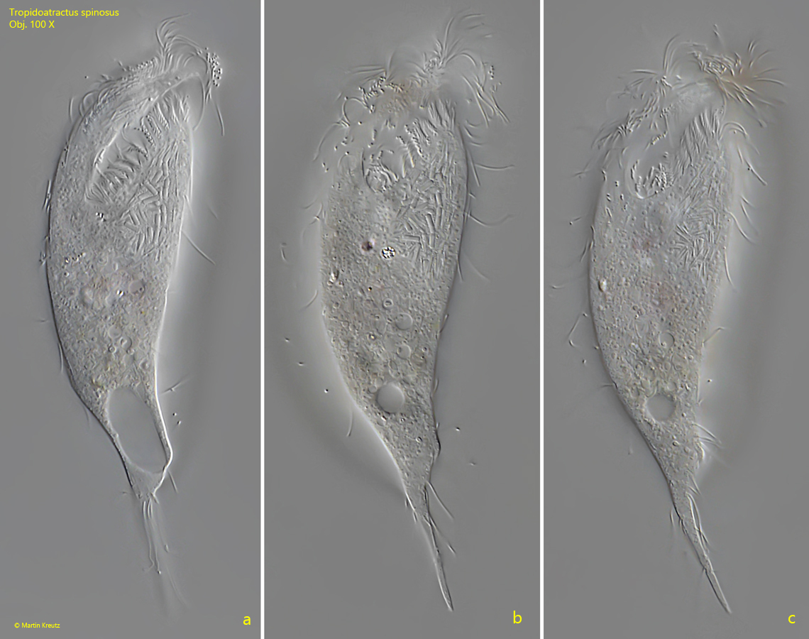
Fig. 2 a-c:Tropidoatractus spinosus. L = 102 µm. Ventral view of a second, freely swimming specimen. The caudal spine of this specimen is also oriented on the right margin of the body. Obj. 100 X.
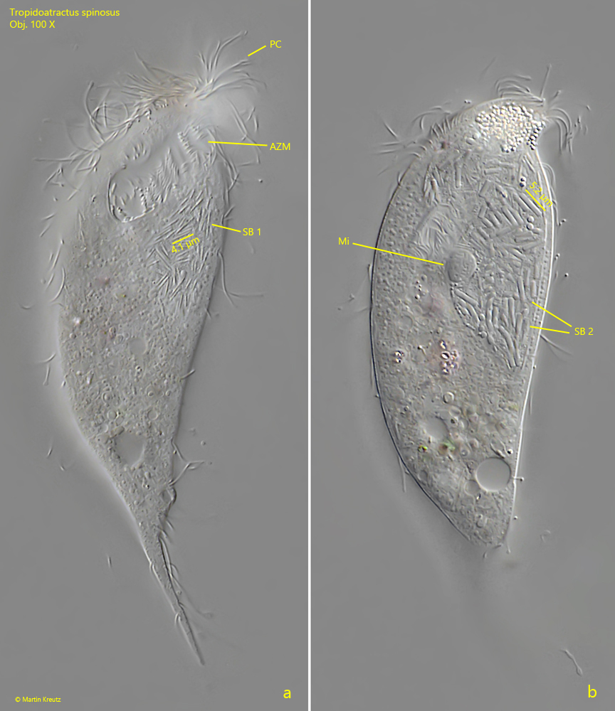
Fig. 3 a-b:Tropidoatractus spinosus. L = 102 µm. Two focal planes on the symbiotic bacteria (SB 1, SB 2) located in the macronucleus. Obviously, thin bacteria with a length of about 4 µm are localized in the marginal region of the macronucleus (SB 1), whereas thicker, rod-shaped bacteria with a length of about 5 µm are found in the center of the macronucleus (SB 2). AZM = adoral zone of membranelles, Mi = micronucleus, PC = perizonal cilia, RG = aggregation of refractive granules in the apical dome. Obj. 100 X.
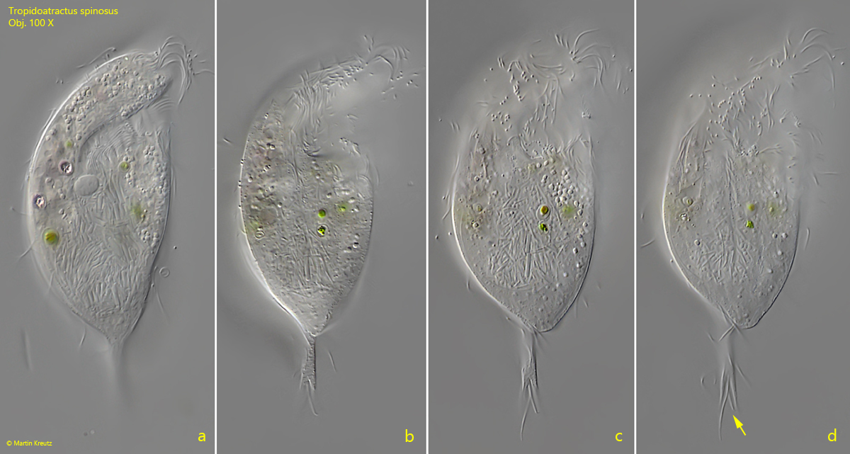
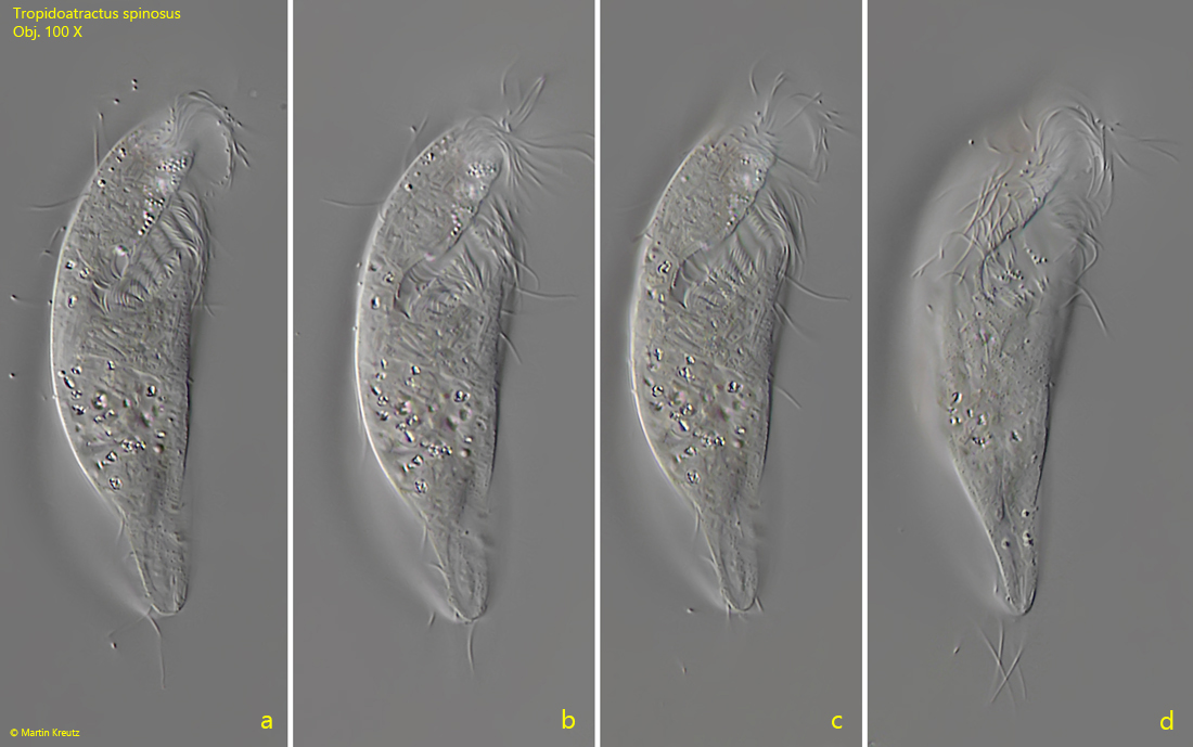
In my population of Tropidoatractusspinosus I also found plumper specimens with a split caudal spine (s. fig. 4 a-d) as well as specimens lacking a caudal spine (s. fig. 5 a-d).
Fig. 4 a-d:Tropidoatractus spinosus. L = 75 µm. A plumper specimen with a split spine (arrow, d). In this specimen, the symbiotic bacteria are also localized in the macronucleus. Obj. 100 X.
Fig. 5 a-d:Tropidoatractus spinosus. L = 64 µm. A freely swimming specimen lacking a caudal spine. Obj. 100 X.