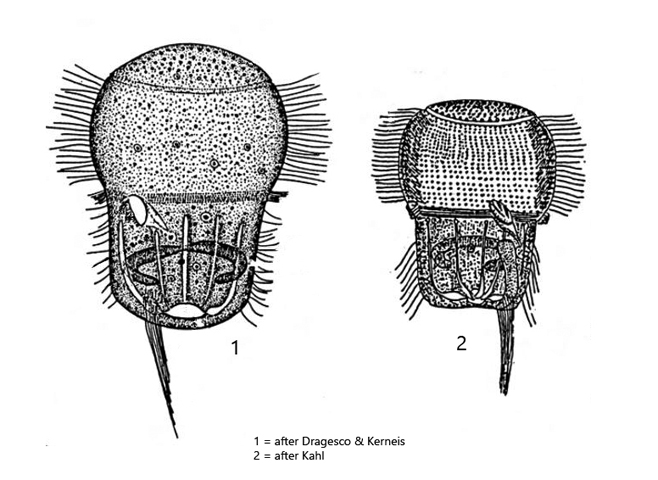
cell barrel-shaped with broadly rounded ends, narrowed in the middle
length 40–110 µm (mostly 50–80 µm)
macronucleus horseshoe-shaped in posterior half
spherical micronucleus
contractile vacuole terminal, with 8–12 collecting canals
fringe of numerous spindle-shaped extrusomes
mouth opening in an equatorial groove
from mouth opening a ciliated groove is running to posterior end
broad girdle of cilia in anterior half
an equatorial girdle of cilia running to the mouth opening
a distinct caudal cirrus of twisted, long cilia
Urocentrum turbo
Urocentrum turbo belongs to the most common ciliates and I find it practically in all my sites. Typical for this ciliate is its gyrating, fast swimming. Via the twisted caudal cilia (s. figs. 2 a and 5) the ciliate can secrete a mucus thread with the help of which it can attach itself to substrates. The thread remains invisible even at high magnification. It can be detached at any time or in case of disturbance, whereupon Urocentrum turbo swims away at great speed. Photographing of free swimming specimens is therefore not easy.
Urocentrum has a complex ciliature. Dominant are two ciliary bands running around the whole body in the anterior half and an equatorial, narrower band in the middle of the body (s. fig. 6 a-b). The latter lies in a groove and is interrupted by the mouth opening (s. fig. 6). The rotating movement creates a fluid flow around the body, which is directed into the mouth opening. There, food (= bacteria) is sorted out and everything else is discharged via another channel, which runs from the mouth opening to the posterior end.
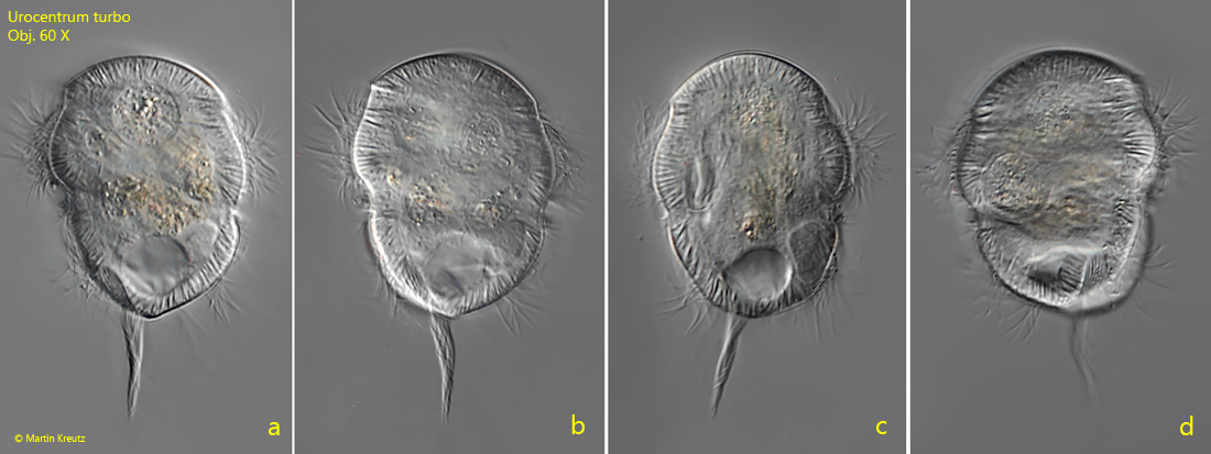
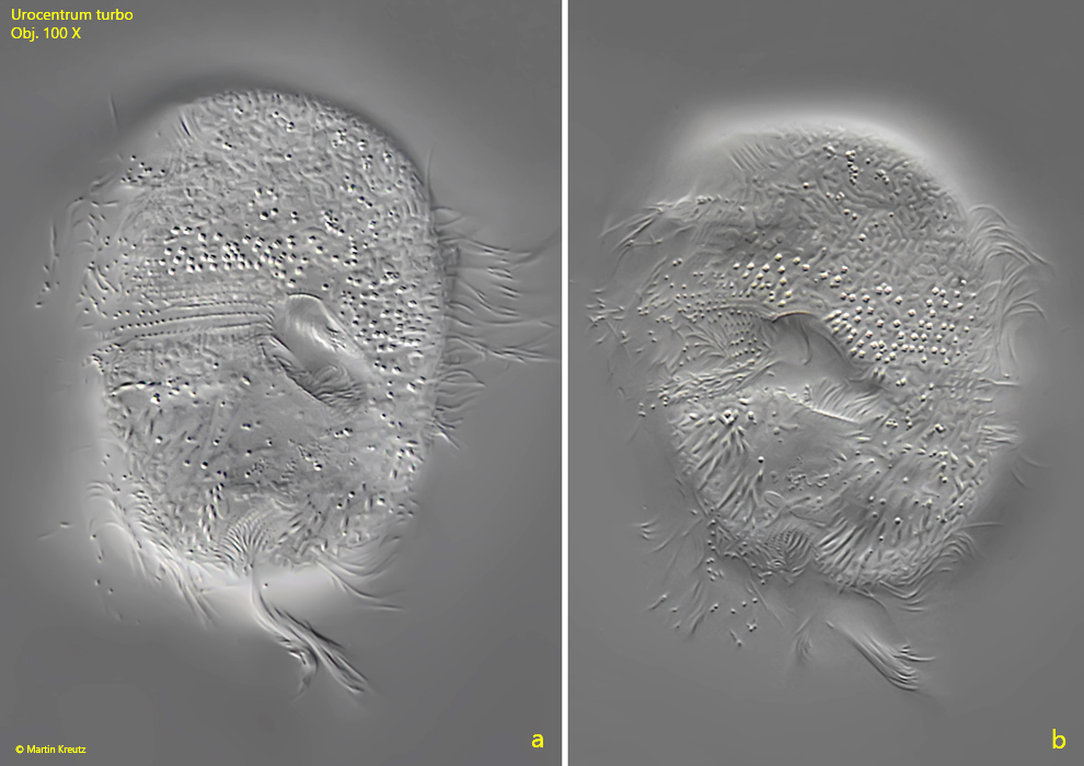
Fig. 1 a-d:Urocentrum turbo. L = 58 µm (without caudal cilia). A freely swimming specimen. Obj. 60 X.
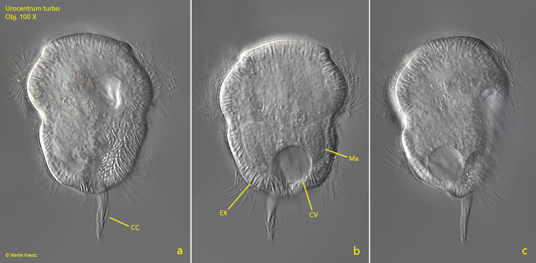
Fig. 2 a-c:Urocentrum turbo. L = 64 µm (without caudal cilia). A second freely swimming specimen. Note the twisted caudal cilia (CC). CV = contractile vacuole, EX = extrusomes, Ma = macronucleus. Obj. 100 X.
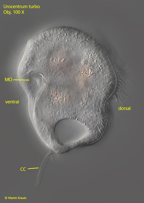
Fig. 3:Urocentrum turbo. L = 58 µm (without caudal cilia). A lateral view from left of a freely swimming specimen. Note the caudal cilia (CC) arising on the ventral side. MO = mouth opening. Obj. 100 X.
Fig. 4 a-b:Urocentrum turbo. L = 55 µm (without caudal cilia). Two focal planes of a ventral view of a slightly squashed specimen gives an impression of the complex ciliation of this ciliate. Obj. 100 X.
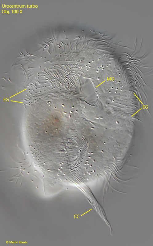
Fig. 5:Urocentrum turbo. L = 55 µm (without caudal cilia). A closer view on the ventral side. The equatorial girdle of cilia (EG) is running to the mouth opening (MO). The long caudal cilia (CC) arise from a vertical girdle below the mouth opening at the posterior end. Obj. 100 X.
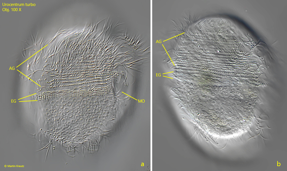
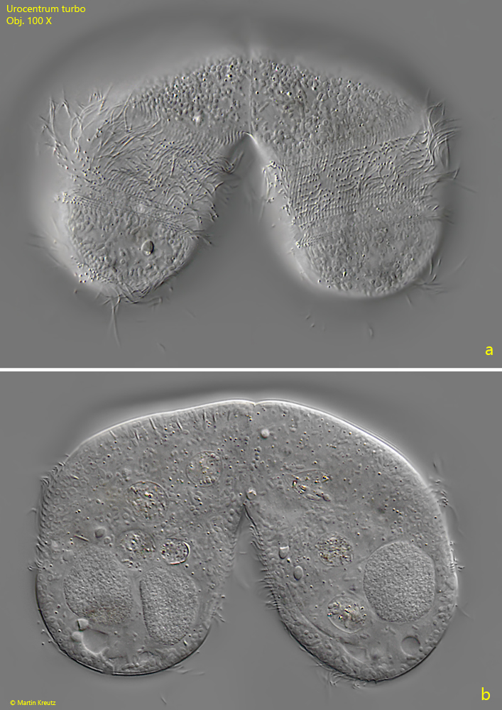
Fig. 6 a-b:Urocentrum turbo. A strongly squashed specimen from right (a) and from dorsal (b). The dense ciliation of the equatorial girdle (EG) is visible and the more spaced rows of the anterior girdle of cilia (AG). MO = mouth opening. Obj. 100 X.
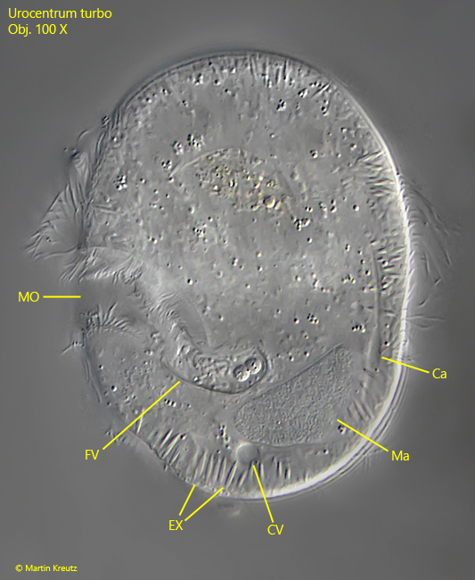
Fig. 7:Urocentrum turbo. A strongly squashed specimen from left with the focal plane on the mouth opening (MO) where a food vacuole is just filled. On the dorsal side one of the collecting canals (Ca) of the contractile vacuole (CV) is visible. Obj. 100 X.
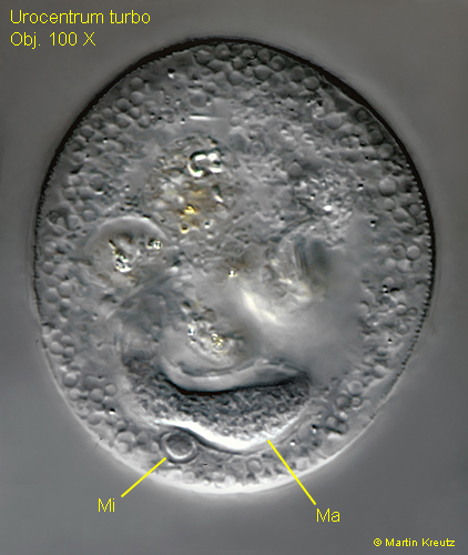
Fig. 8:Urocentrum turbo. A strongly squashed specimen with a visible part of the horseshoe-shaped macronucleus (Ma) and the adjacent spherical micronucleus (Mi). Obj. 100 X.
Fig. 9 a-b:Urocentrum turbo. Two focal planes of a pair in conjugation. Obj. 100 X.