body inversely pear-shaped, posterior end broadly rounded
dome prominent, overhangs the left margin
body dorso-ventrally flattened
adoral zone runs nearly horizontally before it bends almost perpendicular to posterior
length 47–82 µm, width 21–39 µm
distinct fringe of rod-shaped extrusomes beneath pellice, about 2 µm long
cytoplasm with different types of symbiotic bacteria
macronucleus globular or ovoid
one spherical micronucleus adjacent to macronucleus
contractile vacuole terminal
tuft of caudal cilia, about 25 µm long
Urostomides pullus
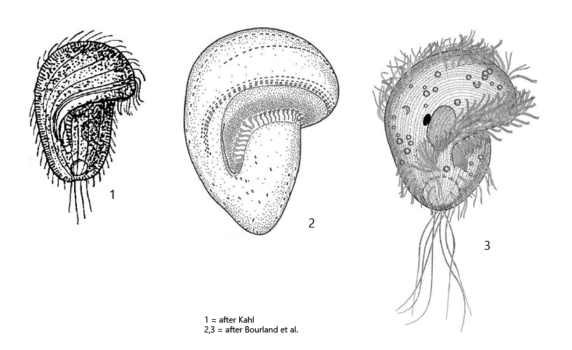
I have found Urostomides pullus only once in Simmelried in January 1996. This metopid ciliate seems to be generally rare. So far Urostomides pullus has only been recorded by Kahl in Germany and by Bourland et al. in the Czech Republic. After 1996 I have no more records of Urostomides pullus in my finding areas.
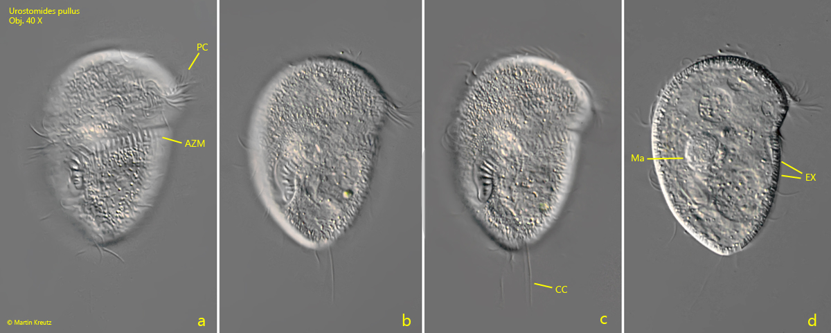
Urostomides pullus can be recognized by the rather stocky body shape and the adoral zone on the ventral side first taking an almost horizontal course before bending almost perpendicular to the posterior end. The mouth opening is located in the posterior third of the body. Characteristic of this species is also the distinct fringe of extrusomes (s. fig. 1d) and the rather long caudal cilia (s. fig. 1c).
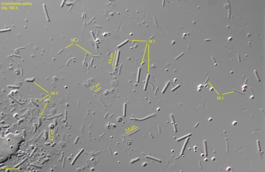
Unfortunately, I only took images with the 40 X lens in 1996 (s. fig. 1 a-d). However, later I squashed the isolated specimen strongly and could identify different forms of bacteria in the cytoplasm, which I consider to be symbiotic bacteria (s. figs. 2 and 3). I could identify at least 4 different types of bacteria:
SB 1 = thick rods with a length of about 6–7 µm
SB 2 = thin rods with a length of about 5–6 µm
SB 3 = short, thin rods with a length of about 2–3 µm
SB 4 = rods with a bent end and a length of about 4–5 µm
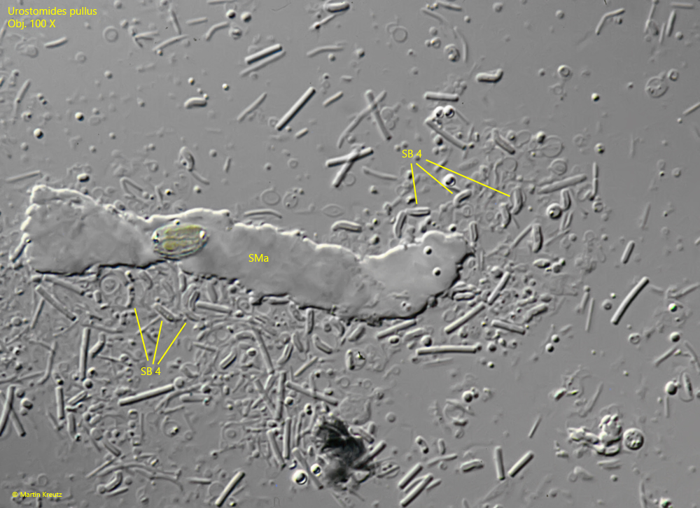
In particular, the bacterial types SB 1 and SB 2 I know well from other anaerobic ciliates. The bacteria of type SB 4 were striking, which not only have a special shape with the bent end, but it also seemed that this type of bacteria are accumulates around the macronucleus. Many bacteria of type SB 4 were found in the strongly squashed specimen around the macronucleus (s. fig. 3). However, confirmation of this assumption can only come from examination of further specimens.
Fig. 1 a-d:Urostomides pullus. L = 75 µm. Ventral view of a freely swimming specimen. AZM = adoral zone of membranelles, CC = caudal cilia, EX = fringe of extrusomes, Ma = macronucleus, PC = perizonal cilia. Obj. 40 X.
Fig. 2:Urostomides pullus. The symbiotic bacteria in the cytoplasm of the strongly squashed specimen shown in fig. 1 a-d. At least 4 types of bacteria are visibe: SB 1 = thick rods with a length of about 6–7 µm, SB 2 = thin rods with a length of about 5–6 µm, SB 3 = short, thin rods with a length of about 2–3 µm, SB 4 = rods with a bent end and a length of about 4–5 µm. Obj. 100 X.
Fig. 3:Urostomides pullus. Another section of the specimen shown in fig. 2. The refractive mass is the squashed macronucleus (SMa). It is noticeable that many SB 4 type bacteria are located around the squashed macronucleus. Possibly this type of bacteria also accumulates around the macronucleus in the living ciliate. Obj. 100 X.