preoral dome broadly rounded, posterior end tapered
length 60–120 µm
cytoplasm colorless, sometimes slightly yellowish
adoral zone runs diagonally around half of the body
posterior sixth of the adoral zone almost parallel to the longitudinal axis of the body
dome overhangs the adoral zone
about 16 ciliary rows
prominent fringe of 2 µm long extrusomes beneath pellice
macronucleus oval or globular, 14–16 µm diameter
micronucleus spherical, 4 µm diameter
contractile vacuole subterminal
tuft of caudal cilia, 15–25 µm long
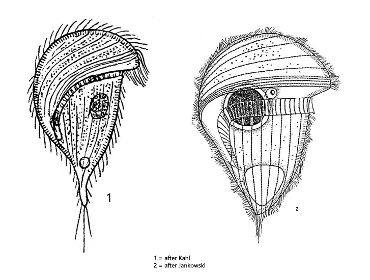
Urostomides striatus
Urostomides striatus was originally described as Metopus striatus. Jankowski then transferred species with 4 perizonal cilia rows to the genus Urostomides. The perizonal cilia rows are difficult to see in vivo, partly because most representatives of this genus are coverslip sensitive, making examination at high magnifications difficult.
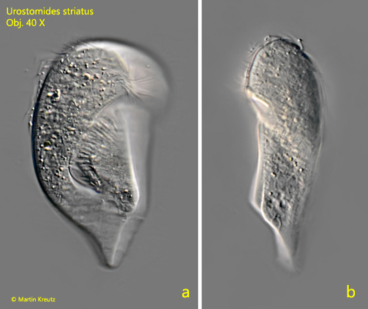
In my site Simmelried I find Urostomides striatus regularly in the upper mud zone. I recognize the species even at low magnifications by the body shape, which resembles a mirror image of a question mark. Also, the body is flattened, but this can only be seen in freely swimming specimens. Urostomides striatus is very coverglass sensitive in my experience. When trying to reduce the layer thickness, the specimens burst and denature immediately. Only in very rare cases careful squashing of specimens succeeds, which made the images with oil immersion fig. 3 a-b and fig. 4 a-b possible.
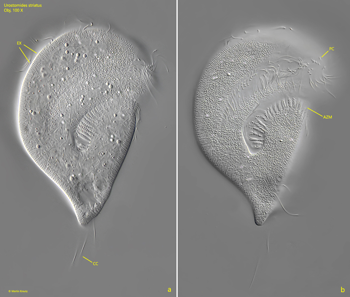
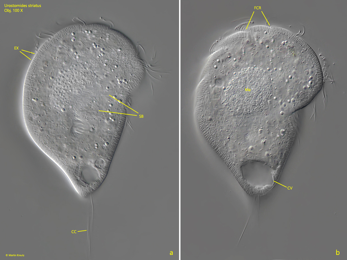
In my population, most specimens were about 80 µm long and possessed a tail-like extended posterior end. The adoral zone extends to about the posterior third of the body. The most caudal section of the adoral zone bends and then runs almost in parallel to the longitudinal axis of the body (s. figs. 2 a, 3 b and 5 b). At high magnifications, the fringe of extrusomes can be clearly seen (s. figs. 3 a and 4 a) and also that symbiotic bacteria are present in the cytoplasm.
Fig. 1 a-b:Urostomides striatus. L = 79 µm. Ventral view (a) and lateral view from right (b). The body ist laterally flattened. Obj. 40 X.
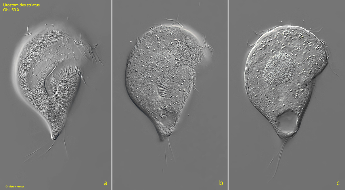
Fig. 2 a-c:Urostomides striatus. L = 84 µm. Ventral view of a second, freely swimming specimen. Obj. 60 X.
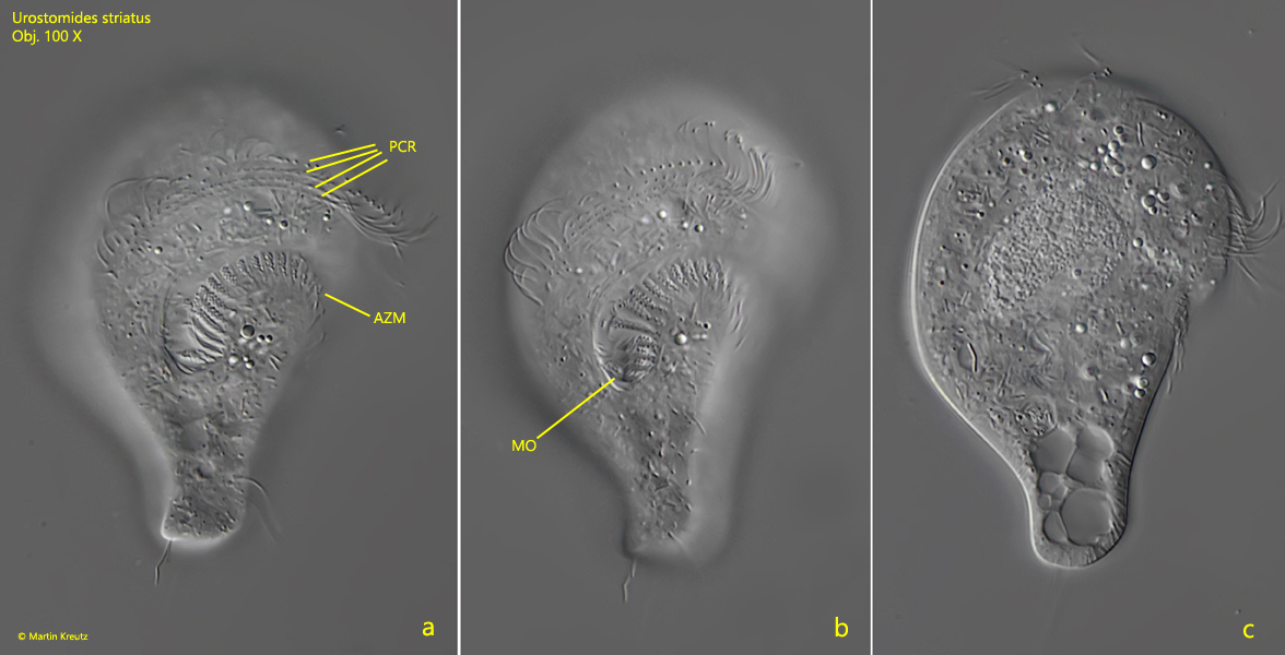
Fig. 3 a-b:Urostomides striatus. L = 84 µm. Two focal planes of the slightly squashed specimen shown in fig. 2 a-c. AZM = adoral zone of membranelles, CC = tuft of caudal cilia, EX = fringe of extrusomes, PC = perizonal cilia. Obj. 100 X.
Fig. 4 a-b:Urostomides striatus. L = 84 µm. Two focal planes of the slightly squashed specimen shown in fig. 2 a-c. CC = tuft of caudal cilia, CV = contractile vacuole, EX = fringe of extrusomes, FCR = furrows between the rows of cilia, Ma = macronucleus, SB = symbiotic bacteria in the cytoplasm. Obj. 100 X.
Fig. 5 a-c:Urostomides striatus. L = 64 µm. Three focal planes of a third, freely swimming specimen in ventral view. Note the 4 rows of perizonal cilia (PCR). AZM = adoral zone of membranelles, MO = mouth opening. Obj. 100 X.