macronucleus globular with an adjacent micronucleus
posterior sixth of body naked
one caudal cilium
movement jumping and spinning
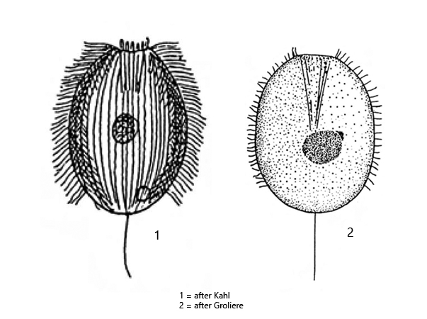
Urotricha ovata
Urotricha ovata is a widespread ciliate, which I could detect in almost all my localities. Especially in old samples with decaying plant masses it is often found.
It is easy to confuse Urotricha ovata with the similar species Urotricha armata. The latter, however, has a dense fringe of extrusomes arranged in parallel to each other. In Urotricha ovata, the extrusomes are scattered under the pellicle and are not arranged in parallel (s. figs. 2 a and 2 b). In addition, the number of longitudinal rows of cilia in Urotricha armata is much higher (35–51) than in Urotricha ovata, where there are only 19–27 rows (s. fig. 1 b).
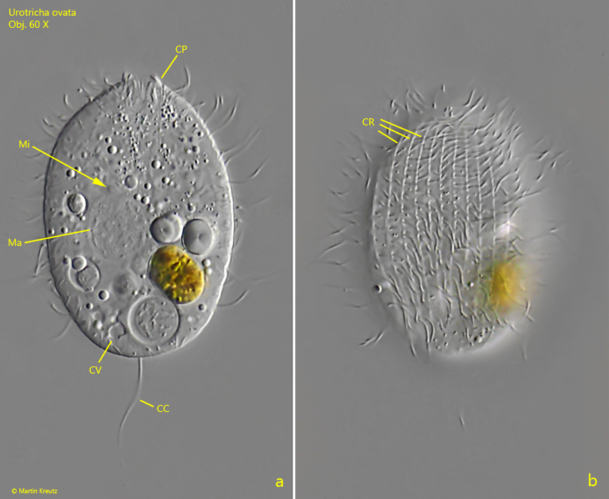
Fig. 1 a-b:Urotricha ovata. L = 43 µm. Two focal planes of a freely swimming specimen. Note the longitudinal rows of cilia (CR). This specimen has 24 rows of cilia. CC = caudal cilium, CV = contractile vacuole, CP = cytopharynx, Ma = macronucleus, Mi = micronucleus. Obj. 60 X.
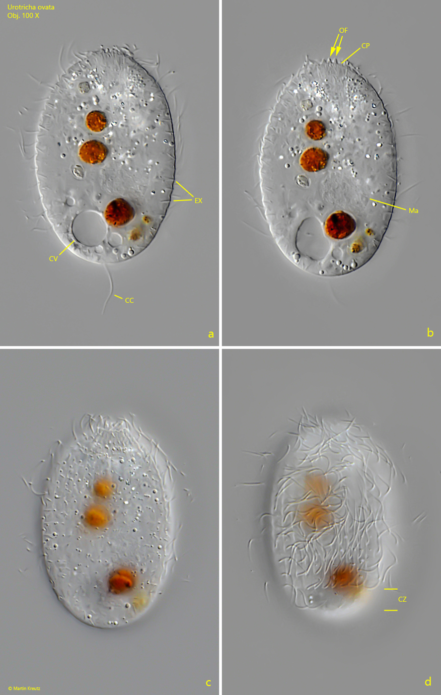
Fig. 2 a-d:Urotricha ovata. L = 43 µm. Four focal planes of a slightly squashed specimen. Note that the posterior sixth of the body is a cilia-free zone (CZ). CC = caudal cilium, CV = contractile vacuole, CP = cytopharynx, EX = extrusomes, Ma = macronucleus, OF = oral flaps. Obj. 100 X.
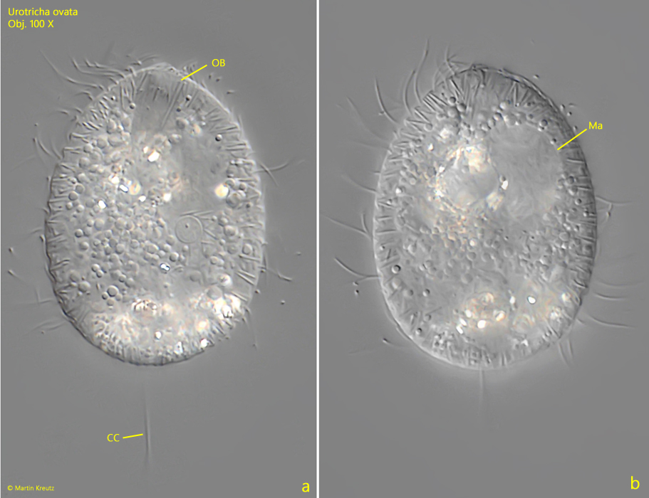
Fig. 3 a-b:Urotricha ovata. L = 42 µm. Two focal planes of a slightly squashed specimen. CC = caudal cilium, Ma = macronucleus, OB = oral basket. Obj. 100 X.